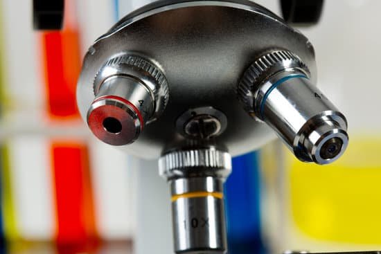
How many microscopes did van leeuwenhoek make? Antonie van Leeuwenhoek made more than 500 optical lenses. He also created at least 25 single-lens microscopes, of differing types, of which only nine have survived. These microscopes were made of silver or copper frames, holding hand-made lenses. Those that have survived are capable of magnification up to 275 times.
How many microscopes did Leeuwenhoek make and what happened to them? Since 1875.
What type of microscope did Leeuwenhoek make? Antonie van Leeuwenhoek used single-lens microscopes, which he made, to make the first observations of bacteria and protozoa.
What 3 things did van Leeuwenhoek discover? As well as being the father of microbiology, van Leeuwenhoek laid the foundations of plant anatomy and became an expert on animal reproduction. He discovered blood cells and microscopic nematodes, and studied the structure of wood and crystals. He also made over 500 microscopes to view specific objects.
How many microscopes did van leeuwenhoek make? – Related Questions
What is the diopter adjustment for on binocular microscopes?
If it focuses at 2 meters, it has a diopter of 1/2. Each microscope eyepiece has a diopter adjustment to allow you to make minor corrections to the image, compensating for the difference in vision between the two eyes.
How to calculate magnification 100x of a microscope?
100X (this means that the image being viewed will appear to be 100 times its actual size). Magnification is not of much value unless resolving power is high.
How many types of microscope are there name them?
There are several different types of microscopes used in light microscopy, and the four most popular types are Compound, Stereo, Digital and the Pocket or handheld microscopes. Some types are best suited for biological applications, where others are best for classroom or personal hobby use.
What if i have microscopic blood in my urine?
Microscopic urinary bleeding is a common symptom of glomerulonephritis, an inflammation of the kidneys’ filtering system. Glomerulonephritis may be part of a systemic disease, such as diabetes, or it can occur on its own.
How to focus 40x microscope?
Using coarse knob, focus using the 10x objective. Then adjust with FINE FOCUS knob on SMALLEST detail visible in the field. Now position 40x objective & adjust FINE FOCUS knob – you SHOULD NOT NEED TO TOUCH COARSE knob. First – close LEFT EYE – then focus the image for your RIGHT EYE using fine focus knob.
Is microscopic hematuria common?
Microscopic hematuria, a common finding on routine urinalysis of adults, is clinically significant when three to five red blood cells per high-power field are visible. Etiologies of microscopic hematuria range from incidental causes to life-threatening urinary tract neoplasm.
Are bed bug eggs microscopic?
They appear like tiny grains of pepper and you can only see the eggs or other parts of their body by looking at them under a microscope. Bed bug larvae actually go through five stages of development.
How to determine total magnification on a microscope?
The total magnification of the microscope is calculated from the magnifying power of the objective multiplied by the magnification of the eyepiece and, where applicable, multiplied by intermediate magnifications.
Is there microscopic life on the moon?
mitis samples found on the camera had indeed survived for nearly three years on the Moon. The paper concluded that the presence of microbes could more likely be attributed to poor clean room conditions rather than the survival of bacteria for three years in the harsh Moon environment.
Can you see a hydrogen atom with a microscope?
Physicists in the US claim to have used a transmission electron microscope (TEM) to see a single hydrogen atom – the first time that a TEM has been used to image such a light atom.
What does urinalysis reflex microscopic urine mean?
This test looks at a sample of your urine under a microscope. It can see cells from your urinary tract, blood cells, crystals, bacteria, parasites, and cells from tumors. This test is often used to confirm the findings of other tests or add information to a diagnosis.
What is low power on a microscope?
Low power objectives cover a wide field of view and they are useful for examining large specimens or surveying many smaller specimens. This objective is useful for aligning the microscope. The power for the low objective is 10X. Place one of the prepared slides onto the stage of your microscope.
What type of microscope is needed to view bacteria?
The compound microscope can be used to view a variety of samples, some of which include: blood cells, cheek cells, parasites, bacteria, algae, tissue, and thin sections of organs. Compound microscopes are used to view samples that can not be seen with the naked eye.
How much can a light microscope magnify up to?
The optical quality of lenses increased and the microscopes are similar to the ones we use today. Throughout their development, the magnification of light microscopes has increased, but very high magnifications are not possible. The maximum magnification with a light microscope is around ×1500.
What is a ferning microscope?
The Claim: In a technique called “ferning,” tiny microscopes are used to examine saliva samples for a fern-like pattern caused when estrogen surges just before ovulation.
How to do direct microscopic count?
Direct counting methods include microscopic counts using a hemocytometer or a counting chamber. The hemocytometer works by creating a volumetric grid divided into differently sized cubes for accurately counting the number of particles in a cube and calculating the concentration of the entire sample.
What is an advantage of an light microscope?
Advantage: Light microscopes have high magnification. Electron microscopes are helpful in viewing surface details of a specimen. Disadvantage: Light microscopes can be used only in the presence of light and have lower resolution. Electron microscopes can be used only for viewing ultra-thin specimens.
What is the main resolution of a scanning electron microscope?
Scanning electron microscope (SEM) is one of the most widely used instrumental methods for the examination and analysis of micro- and nanoparticle imaging characterization of solid objects. One of the reasons that SEM is preferred for particle size analysis is due to its resolution of 10 nm, that is, 100 Å.
How does a microscope help in viewing cells?
A cell is the smallest unit of life. Most cells are so tiny that they cannot be seen with the naked eye. Therefore, scientists use microscopes to study cells. Electron microscopes provide higher magnification, higher resolution, and more detail than light microscopes.
What part supports the microscope?
Base – It acts as microscopes support. It also carries microscopic illuminators. Arms – This is the part connecting the base and to the head and the eyepiece tube to the base of the microscope. It gives support to the head of the microscope and it is also used when carrying the microscope.
What does the stage clips do on a microscope?
Stage clips hold the slides in place. If your microscope has a mechanical stage, you will be able to move the slide around by turning two knobs. One moves it left and right, the other moves it up and down.

