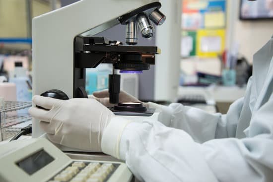
When was the electron microscope created? Ernst Ruska, a German electrical engineer, is credited with inventing the electron microscope. The earliest electron microscope was developed in 1931, and the first commercial, mass-produced instrument became available in 1939.
Who built the electron microscope during 1930s? The transmission electron microscope (TEM) was invented by Ernst Ruska of Germany in the early 1930s, and the first commercial TEM was developed by Siemens in 1939.
Who invented electron microscope in 1940? 1940: Vladimir Zworykin, better known as a co-inventor of television, demonstrates the first electron microscope in the United States.
Who discovered electron microscope in 1935? The scanning electron microscope (SEM) was invented by Max Knoll in 1935, at the Telefunken Company in Berlin, for studying the secondary emission properties of television camera tube targets [1]; four years earlier, he and Ernst Ruska had built the first transmission electron microscope (TEM).
When was the electron microscope created? – Related Questions
Can cells only be seen through a microscope?
“to look at”) to study them. A microscope is an instrument that magnifies objects otherwise too small to be seen, producing an image in which the object appears larger.
What foods to avoid with microscopic colitis?
Avoid beverages that are high in sugar or sorbitol or contain alcohol or caffeine, such as coffee, tea and colas, which may aggravate your symptoms. Choose soft, easy-to-digest foods. These include applesauce, bananas, melons and rice. Avoid high-fiber foods such as beans and nuts, and eat only well-cooked vegetables.
Have we found microscopic life on mars?
To date, no proof of past or present life has been found on Mars. Cumulative evidence suggests that during the ancient Noachian time period, the surface environment of Mars had liquid water and may have been habitable for microorganisms, but habitable conditions do not necessarily indicate life.
Can you see a human egg cell without a microscope?
Most cells aren’t visible to the naked eye: you need a microscope to see them. The human egg cell is an exception, it’s actually the biggest cell in the body and can be seen without a microscope. … That may sound small, but no other cell comes close to being that large.
Why does a microscope slide need to be transparent?
A microscope slide needs to be transparent to allow light to pass through it, which is necessary for magnification to occur.
What can a typical light microscope see?
Light microscopes can be adapted to examine specimens of any size, whole or sectioned, living or dead, wet or dry, hot or cold, and static or fast-moving. They offer a wide range of contrast techniques, providing information on the physical, chemical, and biological attributes of specimens.
What are the lenses called on a microscope?
The compound microscope has two systems of lenses for greater magnification, 1) the ocular, or eyepiece lens that one looks into and 2) the objective lens, or the lens closest to the object. Before purchasing or using a microscope, it is important to know the functions of each part.
How many lenses in a compound microscope?
Typically, a compound microscope is used for viewing samples at high magnification (40 – 1000x), which is achieved by the combined effect of two sets of lenses: the ocular lens (in the eyepiece) and the objective lenses (close to the sample).
What is the use of fine adjustment knob in microscope?
FINE ADJUSTMENT KNOB — A slow but precise control used to fine focus the image when viewing at the higher magnifications.
What is the light source on a microscope used for?
In a modern microscope it consists of a light source, such as an electric lamp or a light-emitting diode, and a lens system forming the condenser. The condenser is placed below the stage and concentrates the light, providing bright, uniform illumination in the region of the object under observation.
When would you need to use a compound microscope?
Typically, a compound microscope is used for viewing samples at high magnification (40 – 1000x), which is achieved by the combined effect of two sets of lenses: the ocular lens (in the eyepiece) and the objective lenses (close to the sample).
What kind of microscope are in classrooms?
The most common types of microscopes used in teaching are monocular light microscopes (80%), followed by binocular optical microscopes (16%), digital microscopes (3%), and stereomicroscopes (1%). A total of 43% of teachers perform microscopy using the demonstration method, and 37% of teachers use practical work.
How to set up microscope scale bar?
In the ‘Analyze/Tools’ menu select ‘Scale Bar’. The scale bar dialog will open and a scale bar will appear on your image. You can adjust the size, color, and placement of your scale bar. Once you are finished click on ‘OK’, save your image, and you are done.
What is a scanning tunneling electron microscope used for?
A scanning tunneling microscope (STM) is a type of microscope used for imaging surfaces at the atomic level. Its development in 1981 earned its inventors, Gerd Binnig and Heinrich Rohrer, then at IBM Zürich, the Nobel Prize in Physics in 1986.
What is the cost of electron microscopes?
The price of a new electron microscope can range from $80,000 to $10,000,000 depending on certain configurations, customizations, components, and resolution, but the average cost of an electron microscope is $294,000. The price of electron microscopes can also vary by type of electron microscope.
What is the term contrast in microscope?
Contrast is defined as the difference in light intensity between the image and the adjacent background relative to the overall background intensity. …
How to count cells with a microscope?
Using a microscope, focus on the grid lines of the counting area with a 5-10x objective. Count the cells in one set of 16 squares (1×1 mm square area; the blue area). You should set a counting rule. To calculate the number of cells per mL: Take the average cell count from each of the sets of 16 corner squares.
What is the function of objective lenses in microscope?
Objective Lenses – The objective lens gathers light from the specimen, magnifies the image of the specimen, and projects the magnified image into the body tube.
What is microscopic characterization?
Microscopy is a category of characterization techniques which probe and map the surface and sub-surface structure of a material. These techniques can use photons, electrons, ions or physical cantilever probes to gather data about a sample’s structure on a range of length scales.
How far can electron microscopes magnify?
This makes electron microscopes more powerful than light microscopes. A light microscope can magnify things up to 2000x, but an electron microscope can magnify between 1 and 50 million times depending on which type you use! To see the results, look at the image below.
What kind of microscope do you need for cell division?
Two types of electron microscopy—transmission and scanning—are widely used to study cells. In principle, transmission electron microscopy is similar to the observation of stained cells with the bright-field light microscope.

