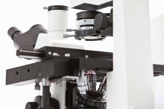
Can bacteria be seen under a microscope? Bacteria are too small to see without the aid of a microscope. While some eucaryotes, such as protozoa, algae and yeast, can be seen at magnifications of 200X-400X, most bacteria can only be seen with 1000X magnification. This requires a 100X oil immersion objective and 10X eyepieces..
What microscope can see bacterial cells? In order to actually see bacteria swimming, you’ll need a lens with at least a 400x magnification. A 1000x magnification can show bacteria in stunning detail. However, at a higher magnification, it can be increasingly difficult to keep them in focus as they move.
How do you identify bacteria under a microscope? Upon viewing the bacteria under the microscope, you will be able to identify the bacteria based on a wide variety of physical characteristics. This mainly involves looking at their shape and size. There are a wide variety of different shapes, yet the three main types are cocci, bacilli, and spiral.
Which microscope is best to see bacteria? On the other hand, compound microscopes are best for looking at all types of microbes down to bacteria. Some, however, are better than others. The magnification for most compound microscopes will be up to 1000X to 2500X.
Can bacteria be seen under a microscope? – Related Questions
Why do we calibrate microscope?
Microscope Calibration can help ensure that the same sample, when assessed with different microscopes, will yield the same results. Even two identical microscopes can have slightly different magnification factors when not calibrated.
What is a scanning objective on a microscope?
A scanning objective lens provides the lowest magnification power of all objective lenses. … The name “scanning” objective lens comes from the fact that they provide observers with about enough magnification for a good overview of the slide, essentially a “scan” of the slide.
Which microscopes can view dna?
To view the DNA as well as a variety of other protein molecules, an electron microscope is used. Whereas the typical light microscope is only limited to a resolution of about 0.25um, the electron microscope is capable of resolutions of about 0.2 nanometers, which makes it possible to view smaller molecules.
Can you see a sperm on a regular microscope?
You can view sperm at 400x magnification. You do NOT want a microscope that advertises anything above 1000x, it is just empty magnification and is unnecessary. In order to examine semen with the microscope you will need depression slides, cover slips, and a biological microscope.
What part of the microscope change magnification?
Revolving Nosepiece or Turret: This is the part of the microscope that holds two or more objective lenses and can be rotated to easily change power (magnification).
What is the function of a compound light microscope?
Typically, a compound microscope is used for viewing samples at high magnification (40 – 1000x), which is achieved by the combined effect of two sets of lenses: the ocular lens (in the eyepiece) and the objective lenses (close to the sample).
How to use fertile focus microscope?
To use Fertile Focus, simply place a drop of saliva from under your tongue on the lens. Once the sample has dried (typically within 5 minutes), press the LED light button and view resultant pattern through the lab-quality magnifying lens.
How does the resolving power of compound microscope?
The resolving power of a compound microscope can be defined as the smallest distance of the object, of which the microscope can form a separate image. … Now resolving power of the compound microscope is given by the reciprocal of the minimum distance between two objects that are to be resolved by the microscope.
What microscope would you use to observe a living organism?
Light microscopes are advantageous for viewing living organisms, but since individual cells are generally transparent, their components are not distinguishable unless they are colored with special stains.
Which microscope is used for viewing living cells?
The light microscope remains a basic tool of cell biologists, with technical improvements allowing the visualization of ever-increasing details of cell structure. Contemporary light microscopes are able to magnify objects up to about a thousand times.
Why is the microscope called a compound light microscope?
The compound light microscope is a tool containing two lenses, which magnify, and a variety of knobs used to move and focus the specimen. Since it uses more than one lens, it is sometimes called the compound microscope in addition to being referred to as being a light microscope.
What does a microscope iris do?
Iris Diaphragm controls the amount of light reaching the specimen. It is located above the condenser and below the stage. Most high quality microscopes include an Abbe condenser with an iris diaphragm. Combined, they control both the focus and quantity of light applied to the specimen.
How does an electron microscope basically work?
The electron microscope uses a beam of electrons and their wave-like characteristics to magnify an object’s image, unlike the optical microscope that uses visible light to magnify images. … This stream is confined and focused using metal apertures and magnetic lenses into a thin, focused, monochromatic beam.
How far can a light microscope magnify?
Throughout their development, the magnification of light microscopes has increased, but very high magnifications are not possible. The maximum magnification with a light microscope is around ×1500.
Why coat gold electron microscope?
It is commonly necessary to coat the sample with a thin layer of gold or gold-palladium alloy in order to prevent charging of the surface, to promote the emission of second- ary electrons so that the specimen conducts evenly, and to provide a homogeneous surface for analysis and imaging.
How to choose a student microscope?
Look for an instrument made of all metal parts without the use of plastic. Ultimately, however, the most important part of a microscope is the lens. Choose the best glass lenses you can afford with total magnification between 40x and 450x. 100x is best for single lens microscopes.
Which lens of a compound microscope?
A compound microscope has multiple lenses: the objective lens (typically 4x, 10x, 40x or 100x) is compounded (multiplied) by the eyepiece lens (typically 10x) to obtain a high magnification of 40x, 100x, 400x and 1000x. Higher magnification is achieved by using two lenses rather than just a single magnifying lens.
Can microscopic colitis turn into cancer?
Microscopic colitis is not related to the more serious types of bowel disease: ulcerative colitis and Crohn’s disease. Microscopic colitis doesn’t make you more likely to get cancer.
How to see scabies without microscope?
Take a dark washable wide-tip marker, and rub around the suspicious bumps or burrows. Then take an alcohol wipe or alcohol-soaked gauze and wipe away the ink. If there’s a scabies burrow under the skin, the ink often remains, showing you a dark irregular line.
How are light microscopes and electron microscopes similar?
Light microscopes and electron microscopes both use radiation – in the form of either light or electron beams, to form larger and more detailed images of objects (e.g. biological specimens, materials, crystal structures, etc.) than the human eye can produce unaided.
How to access the microscope on iphone 7?
On your iPhone or iPad, go to Settings > Accessibility. Tap Magnifier, then turn it on. This adds Magnifier as an accessibility shortcut.

