Can bacteria only be seen with an electron microscope? If you want to look at things like viruses, bacteria, or molecules passing through cell walls, you must use an electron microscope. The devices, developed in the 1930s, use electromagnetic coils to bombard a chemically-prepped, vacuum-sealed specimen with, you guessed it, electrons.
Can you see bacteria with an electron microscope? Scanning electron microscopy allows you to see the outside structure of bacteria in detail (Fig. 3.9) and can be used, for example, to see whether an antimicrobial agent has an impact on cell structure or integrity.
What microbe can only be seen with an electron microscope? chapter 5b
What can an electron microscope see? An electron microscope is a microscope that uses a beam of accelerated electrons as a source of illumination. … Electron microscopes are used to investigate the ultrastructure of a wide range of biological and inorganic specimens including microorganisms, cells, large molecules, biopsy samples, metals, and crystals.
Can bacteria only be seen with an electron microscope? – Related Questions
Why are iodine used under a microscope?
It prevents the slide from drying out when it’s being examined. Iodine stain can be used to stain plant cells to make the internal structures more visible. Most cells are colourless. Stains are used to add contrast.
Can a light microscope see viruses?
Standard light microscopes allow us to see our cells clearly. However, these microscopes are limited by light itself as they cannot show anything smaller than half the wavelength of visible light – and viruses are much smaller than this. But we can use microscopes to see the damage viruses do to our cells.
How hair cuticle looks under microscope?
Under a microscope, human hair looks a lot like animal fur. … Basically, if the scales grow tightly condensed against one another, the hair looks shiny and smooth, but if the scales appear tumbled and disheveled, hair looks unruly and dull.
Can archaea be seen with a light microscope?
However, the archaeal community is still technically limited by what organisms can be cultivated and observed under a microscope.
How to identify fungi under a microscope?
Fungi are identified by their morphology in culture. Fungi have mycelium and spores which are used in the identification. Therefore you have to search for mycelium (hyphae), the spores, origin of the spores, asexual or sexual; and their structure and morphology.
Which describes a compound light microscope?
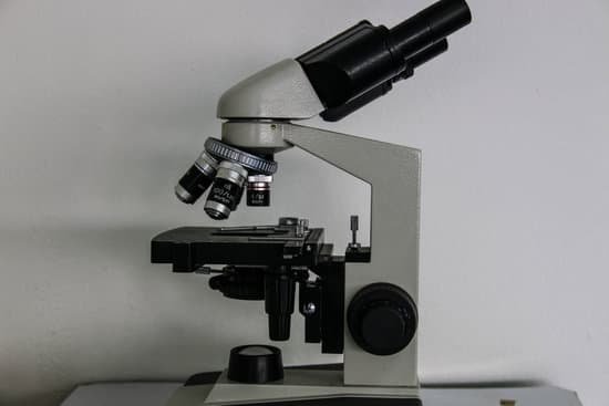
A compound light microscope is a microscope with more than one lens and its own light source. In this type of microscope, there are ocular lenses in the binocular eyepieces and objective lenses in a rotating nosepiece closer to the specimen.
What are microscopic plants?
So microscopic plants refer to plants which consist of a single cell. Microscopic plants are also known as phytoplankton. Complete answer: Algae are single celled mostly aquatic organisms. They belong to the kingdom Protista. The size of algae is microscopic and hence referred to as microscopic plants.
How do you see bacteria under a microscope?
In order to see bacteria, you will need to view them under the magnification of a microscopes as bacteria are too small to be observed by the naked eye. Most bacteria are 0.2 um in diameter and 2-8 um in length with a number of shapes, ranging from spheres to rods and spirals.
How to prevent microscopic colitis symptoms?
There is no way to prevent microscopic colitis, but treating it properly may help prevent the disease from returning. Having microscopic colitis does not increase the chances of getting colon cancer.
What is field of vision on a microscope?
Introduction. Microscope field of view (FOV) is the maximum area visible when looking through the microscope eyepiece (eyepiece FOV) or scientific camera (camera FOV), usually quoted as a diameter measurement (Figure 1).
Can microscopic colitis go away on its own?
Microscopic colitis may get better on its own. But when symptoms persist or are severe, you may need treatment to relieve them.
What is the designation of a compound microscope?
The designation of the microscope as a compound microscope indicates that it has an ocular that focuses on a virtual image of the subject produced in the tube of the microscope by the objective lens. Figure 147. Petrographic microscope.
Who invented the light microscope?
The Dutch spectacle maker Hans Janssen and his son Zacharias are generally credited with creating these compound microscopes. The two of them built what was probably the first compound microscope in the last decade of the 16th century.
Does celiac cause microscopic colitis?
Conclusions: Microscopic colitis is more common in patients with celiac disease than in the general population. Patients with celiac disease and microscopic colitis have more severe villous atrophy and frequently require steroids or immunosuppressant therapies to control diarrhea.
How to adjust resolution on microscope?
The resolution of a specimen viewed through a microscope can be increased by changing the objective lens. The objective lenses are the lenses that protrude downward over the specimen. Grasp the nose piece. The nose piece is the platform on the microscope to which the three or four objective lenses are attached.
What are the different objectives in a compound microscope?
Most compound microscopes come with interchangeable lenses known as objective lenses. Objective lenses come in various magnification powers, with the most common being 4x, 10x, 40x, and 100x, also known as scanning, low power, high power, and (typically) oil immersion objectives, respectively.
What does oil do to a microscope slide?
In light microscopy, oil immersion is a technique used to increase the resolving power of a microscope. This is achieved by immersing both the objective lens and the specimen in a transparent oil of high refractive index, thereby increasing the numerical aperture of the objective lens.
What equipment is used with microscopes?
Optical microscopes use light illumination, lenses, and often a digital camera to magnify images, with a typical resolution limit of about 0.2 µm and magnification in the 1500× range for visible light. Types include stereomicroscopes, compound microscopes, and laser-based Raman systems.
Which type of mirror is used in microscope?
Usually, concave mirror or plano concave mirror are used in microscope. The combination of lenses and mirrors used in making the microscope helps in obtaining magnified and sharp image of the objects.
What microscopic bug is biting me?
Chiggers are tiny parasitic microscopic red bugs that bite humans, birds, and mammals. They’re the larvae of mites belonging to the Trombiculidae family. Chiggers are also known as berry bugs, harvest mites, red bugs, and scrub-itch mites.
What do you see when you look down a microscope?
A microscope lets you look at and study very tiny things in great detail, which the naked eye cannot see. Even under a low-power optical microscope, the fine structures of specimens, or the objects under view, can be seen. Here, the tiny hairs of a stinging nettle are revealed in the microscope view.

