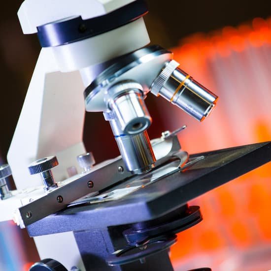
Can you see mono in a microscope? A sample of blood is placed on a microscope slide and mixed with other substances. If heterophil antibodies are present, the blood clumps (agglutinates). This result usually indicates a mono infection.
How can mono be detected? A lot of doctors will do blood tests to confirm mono, though. If someone has symptoms of mono, the doctor may order a complete blood count to look at the lymphocytes, a type of white blood cell that shows specific changes when a person has mono. A doctor may also order a blood test called a monospot.
How long does mono stay on an object? The virus probably survives on an object at least as long as the object remains moist. The first time you get infected with EBV (primary EBV infection) you can spread the virus for weeks and even before you have symptoms. Once the virus is in your body, it stays there in a latent (inactive) state.
What can mono be mistaken for? Mononucleosis is frequently mistaken for other illnesses, such as strep throat, chronic fatigue, or another infection, because the symptoms can overlap, Ramilo says.
Can you see mono in a microscope? – Related Questions
What cell structure cannot be seen with a light microscope?
Some cell parts, including ribosomes, the endoplasmic reticulum, lysosomes, centrioles, and Golgi bodies, cannot be seen with light microscopes because these microscopes cannot achieve a magnification high enough to see these relatively tiny organelles.
How is the magnifying power of the microscope calculated?
To figure the total magnification of an image that you are viewing through the microscope is really quite simple. To get the total magnification take the power of the objective (4X, 10X, 40x) and multiply by the power of the eyepiece, usually 10X.
What does a mechanical stage do on a microscope?
All microscopes are designed to include a stage where the specimen (usually mounted onto a glass slide) is placed for observation. Stages are often equipped with a mechanical device that holds the specimen slide in place and can smoothly translate the slide back and forth as well as from side to side.
What is the use of nosepiece in a microscope?
Nosepiece houses the objectives. The objectives are exposed and are mounted on a rotating turret so that different objectives can be conveniently selected. Standard objectives include 4x, 10x, 40x and 100x although different power objectives are available. Coarse and Fine Focus knobs are used to focus the microscope.
Is aphanitic visible or microscopic?
Small, microscopic, hard-to-see crystals (i.e. no visible crystals) within an igneous rock. This is common in extrusive rocks.
How to focus a microscope on medium power?
To focus a microscope, rotate to the lowest-power objective, and place your sample under the stage clips. Play with the magnification using the coarse adjustment knob and move your slide around until it is centered.
What is the magnification of compound microscope?
Typically, a compound microscope is used for viewing samples at high magnification (40 – 1000x), which is achieved by the combined effect of two sets of lenses: the ocular lens (in the eyepiece) and the objective lenses (close to the sample).
Can you see atoms under electron microscope?
“So we can regularly see single atoms and atomic columns.” That’s because electron microscopes use a beam of electrons rather than photons, as you’d find in a regular light microscope. As electrons have a much shorter wavelength than photons, you can get much greater magnification and better resolution.
How does human hair look under a microscope?
Under a microscope, human hair looks a lot like animal fur. More specifically, it appears as a keratin/ pigment filled tube that’s covered with lots of small external scales. These scales are what tells apart healthy hair from damaged hair.
How is the magnification on a microscope calculated?
The total magnification of the microscope is calculated from the magnifying power of the objective multiplied by the magnification of the eyepiece and, where applicable, multiplied by intermediate magnifications. … If an object is viewed with the eye from a distance of 250 mm, the magnification is 1x.
How is magnification achieved in a microscope?
In simple magnification, light from an object passes through a biconvex lens and is bent (refracted) towards your eye. … Both of these contribute to the magnification of the object. The eyepiece lens usually magnifies 10x, and a typical objective lens magnifies 40x.
What is the function of diaphragm on a microscope?
The field diaphragm controls how much light enters the substage condenser and, consequently, the rest of the microscope.
How does a compound light microscope do?
We shall only learn about the compound light microscope. It uses visible light to visualize the specimen, but passes that light through two separate lens to magnify the image.
How do biological scientist use microscopes?
Cells vary in size. With few exceptions, individual cells are too small to be seen with the naked eye, so scientists use microscopes to study them. A microscope is an instrument that magnifies an object. Most images of cells are taken with a microscope and are called micrographs.
How do the lenses different for compound microscope and electron?
In the case of the compound ones, one lens is placed close to the substance for viewing it. On the other hand, an electron microscope is an instrument that uses electron beams to capture an image and enlarge it.
How does a compound light microscope magnify an image?
A compound microscope uses two or more lenses to produce a magnified image of an object, known as a specimen, placed on a slide (a piece of glass) at the base. … By raising and lowering the stage, you move the lenses closer to or further away from the object you’re examining, adjusting the focus of the image you see.
Do you need a microscope to see a microbe?
In order to see bacteria, you will need to view them under the magnification of a microscopes as bacteria are too small to be observed by the naked eye. … Bacteria have colour only when they are present in a colony, single bacteria are transparent in appearance.
What can you see with a tem microscope?
The transmission electron microscope is used to view thin specimens (tissue sections, molecules, etc) through which electrons can pass generating a projection image. The TEM is analogous in many ways to the conventional (compound) light microscope.
What is a metallurgical microscope used for?
Metallographic microscopes are used to identify defects in metal surfaces, to determine the crystal grain boundaries in metal alloys, and to study rocks and minerals. This type of microscope employs vertical illumination, in which the light source is inserted into the microscope tube…
What sets the limit for resolution on a microscope?
To achieve the maximum (theoretical) resolution in a microscope system, each of the optical components should be of the highest NA available (taking into consideration the angular aperture). In addition, using a shorter wavelength of light to view the specimen will increase the resolution.
What shows hematuria microscopic in urine test?
Microscopic hematuria, a common finding on routine urinalysis of adults, is clinically significant when three to five red blood cells per high-power field are visible. Etiologies of microscopic hematuria range from incidental causes to life-threatening urinary tract neoplasm.

