Can you take a video of electron microscope? These microscopes use a beam of electrons to illuminate extremely small structures, but they can’t properly capture something while it’s moving. With a laser-assisted electron microscope, you can capture a video of something while it’s moving, but they’re extremely expensive.
Can you view live specimen electron microscope? Electron microscopes use a beam of electrons instead of beams or rays of light. Living cells cannot be observed using an electron microscope because samples are placed in a vacuum.
Can electron microscopes take pictures? Scanning electron microscopes (SEMs) use a focused beam of electrons to produce images of objects that have been magnified up to 2,000,000 times, revealing detail and complexity inaccessible with light microscopy.
What is the function of a simple microscope? A simple microscope is used to see the magnified image of an object. Antonie Van Leeuwenhoek, a Dutch, invented the first simple microscope, consisting of a small single high powered converging lens to inspect the small micro-organisms of freshwater. It is chiefly designed from the light microscope.
Can you take a video of electron microscope? – Related Questions
What do modern microscopes use lenses to bend?
A simple light microscope manipulates how light enters the eye using a convex lens, where both sides of the lens are curved outwards. When light reflects off of an object being viewed under the microscope and passes through the lens, it bends towards the eye.
How do microscopes help people?
A microscope lets the user see the tiniest parts of our world: microbes, small structures within larger objects and even the molecules that are the building blocks of all matter. The ability to see otherwise invisible things enriches our lives on many levels.
What is the path of light through the compound microscope?
The path of light through a microscope. Modern microscopes are complex precision instruments. Light, originating in the light source (1), is focused by the condensor (2) onto the specimin (3). The light then enters the objective lens (4) and the image is magnified.
Are nematodes microscopic?
Nematodes range in size from microscopic to 7 metres (about 23 feet) long, the largest being the parasitic forms found in whales. … Nematodes can cause a variety of diseases (such as filariasis, ascariasis, and trichinosis) and parasitize many crop plants and domesticated animals.
What type of microscope is used ca 2 imaging?
In recent years, the number of researchers performing calcium imaging (Ca imaging) using a fluorescence microscope has increased. Calcium imaging is a method in which the flow of intracellular calcium is measured so as to directly observe the calcium signaling of active neurons.
What microscope do you use for gram stain?
Observing a Gram stain in a light microscope. The light microscope is arguably the most valuable research tool in the history of biology.
Who invented the compound light microscope?
The Dutch spectacle maker Hans Janssen and his son Zacharias are generally credited with creating these compound microscopes. The two of them built what was probably the first compound microscope in the last decade of the 16th century.
What is microscopic observation?
LP40224-5 Microscopic observation. Microscopy is a technique that uses microscopes to examine very small objects, not seen by the naked eye. There are three well-known branches of microscopy: optical, electron and scanning probe microscopy.
Why use oil in microscope?
In light microscopy, oil immersion is a technique used to increase the resolving power of a microscope. This is achieved by immersing both the objective lens and the specimen in a transparent oil of high refractive index, thereby increasing the numerical aperture of the objective lens.
What is mirror in microscope?
Mirrors in the microscope’s interior are used to focus light to make the microscope more compact, or to make it easier to make the microscope binocular. On low-cost compound microscopes, the mirror is used to focus light from underneath the slide through the microscope’s objective lens.
How to get rid of microscopic hematuria?
Depending on the condition causing your hematuria, treatment might involve taking antibiotics to clear a urinary tract infection, trying a prescription medication to shrink an enlarged prostate or having shock wave therapy to break up bladder or kidney stones. In some cases, no treatment is necessary.
What part of the microscope is used to focus?
Coarse Adjustment Knob- The coarse adjustment knob located on the arm of the microscope moves the stage up and down to bring the specimen into focus. The gearing mechanism of the adjustment produces a large vertical movement of the stage with only a partial revolution of the knob.
Can drinking cause microscopic hematuria?
Can Alcohol Use Lead to Blood in Urine? Alcohol is not typically a direct cause of blood in urine. However, that is not to say it doesn’t contribute to other conditions that may cause blood in urine. If long-term alcohol use occurs, it can damage the kidneys, which may cause blood in urine.
What is the maximum magnification possible with a light microscope?
Using the mathematical equations given above and the values for maximum numerical aperture attainable with the lenses of a light microscope it can be shown that the maximum useful magnification on a light microscope is between 1000X and 1500X. Higher magnification is possible, but resolution will not improve.
What is the lowest magnification for the compound light microscope?
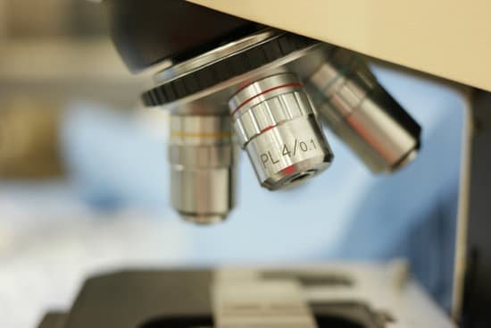
Why do you need to start with 4x in magnification on a microscope? The 4x objective lens has the lowest power and, therefore the highest field of view. As a result, it is easier to locate the specimen on the slide than if you start with a higher power objective.
What microscope would you use to examine bullet casings?
Stereo microscopes are used to determine basic class characteristics of fired bullets, bullet fragments and cartridge/shotshell cases. A comparison microscope is used for the examination of fired bullets, bullet fragments and cartridge/shotshell cases.
When robert hooke invented the microscope?
During this period, Hooke’s interest in microscopy and astronomy soared, and he published Micrographia, his best known work on optical microscopy in 1665. The next year, Hooke published a volume on comets, Cometa, detailing his close observation of the comets occurring in 1664 and 1665.
What is the meaning of compound microscope?
A compound microscope is a microscope that uses multiple lenses to enlarge the image of a sample. … Compound microscopes usually include exchangeable objective lenses with different magnifications (e.g 4x, 10x, 40x and 60x), mounted on a turret, to adjust the magnification.
Where was the compound microscope invented?
Janssen was the son of a spectacle maker named Hans Janssen, in Middleburg, Holland, and while Zacharias is credited with inventing the compound microscope, most historians surmise that his father must have played a vital role, since Zacharias was still in his teens in the 1590s.

