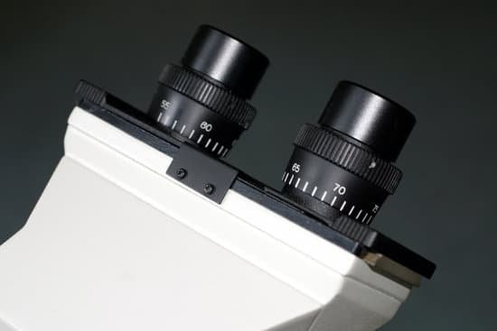
Can you use alcohol to clean a microscope? Alcohols are the most common type of disinfectant and are safe to use on most microscope components except for materials like rubber or specific plastics. For optical components like eyepieces, condensers, and objectives, 70% ethanol is safe to use.
Can you clean a microscope with alcohol? As you clean and disinfect your microscope, always follow proper hand hygiene. … If soap and water aren’t available and your hands don’t look noticeably dirty, then use an alcohol-based hand sanitizer that contains at least 60% alcohol.
Why is alcohol not used to clean microscope? Do not use alcohol, acetone or other solvents on your instrument as they may cause damage to painted surfaces. For glass stage plates, we recommend using mild soapy water and using your bare hands. Have a soft paper or cloth towel to set the glass on after you’ve washed and rinsed your glass.
How do you properly clean a microscope? To clean microscope eyepiece lenses, breathe condensation onto them and then wipe them with lens tissue. Kim-wipes are made by Kleenex and generally will work well. For stubborn spots, wipe the surface with tissue moistened with 95% alcohol. Wipe the lens dry with a dry tissue.
Can you use alcohol to clean a microscope? – Related Questions
Is neon microscopic or macroscopic?
We are given an electronic configuration for the Neon atom. Atoms are not seen with the naked eye. Therefore, an electronic configuration is a microscopic property.
How to use a monocular microscope?
The proper way to use a monocular microscope is to look through the eyepiece with one eye and keep the other eye open (this helps avoid eye strain). Remember, everything is upside down and backwards. When you move the slide to the right, the image goes to the left!
What is the strongest microscope lens you can buy?
The oil immersion objective lens provides the most powerful magnification, with a whopping magnification total of 1000x when combined with a 10x eyepiece.
What is the compound light microscope field of view?
Typically, a compound microscope is used for viewing samples at high magnification (40 – 1000x), which is achieved by the combined effect of two sets of lenses: the ocular lens (in the eyepiece) and the objective lenses (close to the sample).
Is cytoskeleton visible under light microscope?
These proteins can be used for passing messages (hormones) and repairing damage (cytoskeleton). This structure is too small to be seen by the average light microscope.
What is a transmission electron microscope good for?
Transmission electron microscopy is a major analytical method in the physical, chemical and biological sciences. TEMs find application in cancer research, virology, and materials science as well as pollution, nanotechnology and semiconductor research, but also in other fields such as paleontology and palynology.
How to remove fungus from microscope lens?
A hydrogen peroxide blend with ammonia is a good method, as is a vinegar and water solution to remedy the fungus problem. Make sure you don’t delay, or you’ll need to have the lens professionally dismantled and cleaned, which will be expensive.
How to identify tissues under a microscope?
There are three basic shapes used to classify epithelial cells. A squamous epithelial cell looks flat under a microscope. A cuboidal epithelial cell looks close to a square. A columnar epithelial cell looks like a column or a tall rectangle.
How to slow down protozoa under the microscope?
The idea here is to thicken the water in which the microscopic animals are moving. Glue-like substances, such as gum Arabic, gum tragacanth, unflavored gelatin, methylcellulose, or carboxymethylcellulose can be added to the microscope slide preparation. Proprietary slowing agents may be solutions of methylcellulose.
What is the purpose of the diaphragm on a microscope?
Field planes are controlled via the field diaphragm. The field diaphragm in the base of the microscope controls only the width of the bundle of light rays reaching the condenser. This variable aperture does not affect the optical resolution, numerical aperture, or the intensity of illumination.
What are two basic morphological types of microscopic fungi?
Fungi can be divided into two basic morphological forms, yeasts and hyphae. Yeastsare unicellular fungi which reproduce asexually by blastoconidia formation (budding) or fission.
How to clean microscope slide?
When slides get soiled, you can clean them with soapy water or isopropyl alcohol. Do not immerse slides in water or soak them in it. This loosens the cover glass adhesive, causing the cover glass to come off and possibly ruin the slide.
Can a microscope laser burn paper?
A Class 3B laser is hazardous if the eye is exposed directly, but diffuse reflections such as those from paper or other matte surfaces are not harmful. The AEL for continuous lasers in the wavelength range from 315 nm to far infrared is 0.5 W.
How much blood to put of a microscope slide?
Place a drop of blood approximately 4 mm in diameter on the slide (near the end if one smear is to be made, or at the proper location if two smears are to share a slide). See the drawing below.
What is piezoelectric scanner scanning probe microscope?
Scanning probe microscopes are a family of instruments used for studying surface properties of materials from the micrometer all the way down to the atomic level. … The scanner controls the precise position of the probe in relation to the surface, both vertically and laterally.
What is a unicellular organism under a microscope?
Unicellular organisms are made up of only one cell that carries out all of the functions needed by the organism, while multicellular organisms use many different cells to function. Unicellular organisms include bacteria, protists, and yeast.
What is the overall magnification of this microscope?
Total Magnification: To figure the total magnification of an image that you are viewing through the microscope is really quite simple. To get the total magnification take the power of the objective (4X, 10X, 40x) and multiply by the power of the eyepiece, usually 10X.
Why are microscopes important to understanding cell biology?
Because most cells are too small to be seen by the naked eye, the study of cells has depended heavily on the use of microscopes. … Thus, the cell achieved its current recognition as the fundamental unit of all living organisms because of observations made with the light microscope.
What type of lenses are used in a compound microscope?
A compound microscope is made of two convex lenses; the first, the ocular lens, is close to the eye, and the second is the objective lens.
Can light microscopes see live bacteria?
Generally speaking, it is theoretically and practically possible to see living and unstained bacteria with compound light microscopes, including those microscopes which are used for educational purposes in schools.
What date did zacharias janssen invent the microscope?
In Boreel’s investigation Johannes also claimed his father, Zacharias Janssen, invented the compound microscope in 1590. For this to be true (Zacharias most likely dates of birth would have made him 2–5 years old at the time) some historians concluded grandfather Hans Martens must have invented it.

