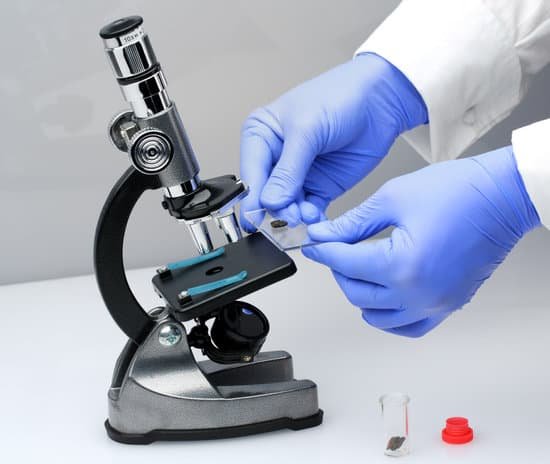
Did robert hooke invent the light microscope? Hooke was one of a small handful of scientists to embrace the first microscopes, improve them, and use them to discover nature’s hidden details. He designed his own light microscope, which used multiple glass lenses to light and magnify specimens.
Who first invented light microscope? 1590: Two Dutch spectacle-makers and father-and-son team, Hans and Zacharias Janssen, create the first microscope.
Did Hooke invent the microscope? Although Hooke did not make his own microscopes, he was heavily involved with the overall design and optical characteristics. The microscopes were actually made by London instrument maker Christopher Cock, who enjoyed a great deal of success due to the popularity of this microscope design and Hooke’s book.
What was Hooke most famous for? English physicist Robert Hooke is known for his discovery of the law of elasticity (Hooke’s law), for his first use of the word cell in the sense of a basic unit of organisms (describing the microscopic cavities in cork), and for his studies of microscopic fossils, which made him an early proponent of a theory of …
Did robert hooke invent the light microscope? – Related Questions
What microscope magnifies objects in steps?
What is a compound light microscope? an instrument that uses light and a series of lenses to magnify objects in steps.
What color is copper under a microscope?
Copper has the chemical element symbol Cu and atomic number 29. Copper is a highly thermal and electrically conducive metal. Pure copper is soft and quite malleable, usually with a reddish orange color.
Can lysosomes be seen with a light microscope?
Lysosomes/Endosome. Again, individual endosomes and lysosomes are not visible using regular light microscopy. However, in some cell types, such as macrophages, these cellular compartments show up in regular histological sections as granular inclusions in the cytoplasm.
Who created the sem microscope?
In 1937, Bodo von Borries and Helmut Ruska joined him to develop ways that the principles could be applied, such as to examine biological samples. In the same year, Manfred von Ardenne developed the first scanning electron microscope.
How to oil immersion microscope?
To use an oil immersion lens, first focus on the area of specimen to be observed with the high dry (400x) lens. Place a drop of immersion oil on the cover slip over that area, and very carefully swing the oil immersion lens into place.
Where is the electron microscope used?
Electron microscopes are used to investigate the ultrastructure of a wide range of biological and inorganic specimens including microorganisms, cells, large molecules, biopsy samples, metals, and crystals. Industrially, electron microscopes are often used for quality control and failure analysis.
How small can the atomic force microscope clearly see?
The smallest thing that we can see with a ‘light’ microscope is about 500 nanometers. One nanometer is one-billionth (that’s 1,000,000,000th) of a meter. So the smallest thing that you can see with a light microscope is about 200 times smaller than the width of a human hair. Bacteria are about 1000 nanometers in size.
What is a monocular compound microscope?
The basic form of a compound microscope is monocular: a single tube is used, with the objective at one end and a single eyepiece at the other. … A true stereoscopic microscope is configured by using two objectives and two eyepieces, enabling each eye to view the object separately, making it appear three-dimensional.
How do you find total magnification of a microscope?
The total magnification of the microscope is calculated from the magnifying power of the objective multiplied by the magnification of the eyepiece and, where applicable, multiplied by intermediate magnifications. A distinction is made between magnification and lateral magnification.
How to hold and use a microscope?
Hold the microscope with one hand around the arm of the device, and the other hand under the base. This is the most secure way to hold and walk with the microscope. Avoid touching the lenses of the microscope. The oil and dirt on your fingers can scratch the glass.
Who was the first person to discover microscopic organisms?
The existence of microscopic organisms was discovered during the period 1665-83 by two Fellows of The Royal Society, Robert Hooke and Antoni van Leeuwenhoek. In Micrographia (1665), Hooke presented the first published depiction of a microganism, the microfungus Mucor.
When microscope not in focus?
The height of your condenser may be set too high or too low (this can also affect resolution). Make sure that your objective lenses are screwed all the way into the body of the microscope. On high school microscopes, if someone adjusts the rack stop, the microscope will not focus.
Which two parts are used when carrying the microscope?
When carrying a compound microscope always take care to lift it by both the arm and base, simultaneously. There are two optical systems in a compound microscope: Eyepiece Lenses and Objective Lenses: Eyepiece or Ocular is what you look through at the top of the microscope.
What is the formula for total magnification on a microscope?
To calculate total magnification, find the magnification of both the eyepiece and the objective lenses. The common ocular magnifies ten times, marked as 10x. The standard objective lenses magnify 4x, 10x and 40x. If the microscope has a fourth objective lens, the magnification will most likely be 100x.
What is difference between macroscopic and microscopic?
The macroscopic level includes anything seen with the naked eye and the microscopic level includes atoms and molecules, things not seen with the naked eye. Both levels describe matter.
Why do images in microscopes flip?
The eyepiece of the microscope contains a 10x magnifying lens, so the 10x objective lens actually magnifies 100 times and the 40x objective lens magnifies 400 times. There are also mirrors in the microscope, which cause images to appear upside down and backwards.
What is the difference between gross and microscopic anatomy?
“Gross anatomy” customarily refers to the study of those body structures large enough to be examined without the help of magnifying devices, while microscopic anatomy is concerned with the study of structural units small enough to be seen only with a light microscope. Dissection is basic to all anatomical research.
What is the function of a condenser in a microscope?
On upright microscopes, the condenser is located beneath the stage and serves to gather wavefronts from the microscope light source and concentrate them into a cone of light that illuminates the specimen with uniform intensity over the entire viewfield.
How many people have microscopic colitis?
Microscopic colitis can develop at any time, but it is more common in middle-age, with those affected often diagnosed between the ages of 50 and 70. It also occurs more frequently in women and can occur earlier in people who smoke. Microscopic colitis occurs in 18 people in 100,000 people, per year.
Which is a benefit of the light microscope?
Advantage: Light microscopes have high magnification. Electron microscopes are helpful in viewing surface details of a specimen. Disadvantage: Light microscopes can be used only in the presence of light and have lower resolution.
How to calculate total magnification in a microscope?
To figure the total magnification of an image that you are viewing through the microscope is really quite simple. To get the total magnification take the power of the objective (4X, 10X, 40x) and multiply by the power of the eyepiece, usually 10X.

