Do light microscopes have better resolution than electron? Electrons have much a shorter wavelength than visible light, and this allows electron microscopes to produce higher-resolution images than standard light microscopes.
How is resolution different in light and electron microscopes? There are two main types of microscope: light microscopes are used to study living cells and for regular use when relatively low magnification and resolution is enough. electron microscopes provide higher magnifications and higher resolution images but cannot be used to view living cells.
Which type of microscope has the best resolution? The microscope that can achieve the highest magnification and greatest resolution is the electron microscope, which is an optical instrument that is designed to enable us to see microscopic details down to the atomic scale (check also atom microscopy).
Is a light microscope high resolution? light microscopes are used to study living cells and for regular use when relatively low magnification and resolution is enough. electron microscopes provide higher magnifications and higher resolution images but cannot be used to view living cells.
Do light microscopes have better resolution than electron? – Related Questions
What is the function of microscope parts?
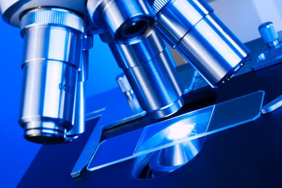
All of the parts of a microscope work together – The light from the illuminator passes through the aperture, through the slide, and through the objective lens, where the image of the specimen is magnified.
What can be determined by the microscopic evaluation of hair?
Microscopic examination of hair can determine. … DNA may be found in the hair root and can be used to determine individual characteristics.
What is the iris on a microscope?
Iris Diaphragm controls the amount of light reaching the specimen. It is located above the condenser and below the stage. Most high quality microscopes include an Abbe condenser with an iris diaphragm. Combined, they control both the focus and quantity of light applied to the specimen.
How many lenses does the light pass through a microscope?
Ordinary light microscopes are equipped with three objective lenses (5 ×/10 ×, 40 ×, and 90/100 ×), and two ocular (5 ×, 10 ×) lenses.
Which part of the microscope concentrates light through the specimen?
Condenser: Concentrates the light on the specimen. The condenser has a height- adjustment knob that raises and lowers the condenser to vary light delivery. Generally, the best position for the condenser is close to the inferior surface of the stage.
What kind of glass are used with microscopes?
Microscope slides are usually made of optical quality glass, such as soda lime glass or borosilicate glass, but specialty plastics are also used.
What does the coarse arm do on the microscope?
Coarse Adjustment Knob- The coarse adjustment knob located on the arm of the microscope moves the stage up and down to bring the specimen into focus.
What allows us to view flagella under the microscope?
The flagella stain allows observation of bacterial flagella under the light microscope. Bacterial flagella are normally too thin to be seen under such conditions.
Which of the following microscopes has the highest resolution?
Out of all types of microscopes, the electron microscope has the greatest capability in achieving high magnification and resolution levels, enabling us to look at things right down to each individual atom.
What does resolution mean in microscopes?
In microscopy, the term ‘resolution’ is used to describe the ability of a microscope to distinguish detail. In other words, this is the minimum distance at which two distinct points of a specimen can still be seen – either by the observer or the microscope camera – as separate entities.
What year was the comparison microscope invented?
History. One of the first prototypes of a comparison microscope was developed in 1913 in Germany. In 1929, using a comparison microscope adapted for forensic ballistics, Calvin Goddard and his partner Phillip Gravelle were able to absolve the Chicago Police Department of participation in the St.
When were transmission electron microscopes invented?
Ernst Ruska at the University of Berlin, along with Max Knoll, combined these characteristics and built the first transmission electron microscope (TEM) in 1931, for which Ruska was awarded the Nobel Prize for Physics in 1986.
How to make a smartphone microscope?
Cut two small strips of plastic to create a microscope slide, and place a droplet of water from a puddle between the strips. Place the slide on the white paper on top of a flashlight. Focus your DIY phone microscope, and zoom in on the sample until you see organisms.
What part of the microscope controls the amount of light?
Iris diaphragm dial: Dial attached to the condenser that regulates the amount of light passing through the condenser. The iris diaphragm permits the best possible contrast when viewing the specimen.
What does the coarse focus do on a microscope?
COARSE ADJUSTMENT KNOB — A rapid control which allows for quick focusing by moving the objective lens or stage up and down. It is used for initial focusing.
What is a binocular dissecting microscope?
While a dissecting microscope has a binocular eyepiece like a compound microscope, the image you get from a stereo microscope is, like its name implies, stereoscopic: you get an offset image in each eyepiece to create a three-dimensional image with a discernible depth of field. …
What year was the first microscope made?
Lens Crafters Circa 1590: Invention of the Microscope. Every major field of science has benefited from the use of some form of microscope, an invention that dates back to the late 16th century and a modest Dutch eyeglass maker named Zacharias Janssen.
How to calculate eyepiece magnification microscope?
To figure the total magnification of an image that you are viewing through the microscope is really quite simple. To get the total magnification take the power of the objective (4X, 10X, 40x) and multiply by the power of the eyepiece, usually 10X.
What is an electron microscope used to see?
Electron microscopes are used to investigate the ultrastructure of a wide range of biological and inorganic specimens including microorganisms, cells, large molecules, biopsy samples, metals, and crystals. Industrially, electron microscopes are often used for quality control and failure analysis.
What does a modern light microscope do?
A simple light microscope manipulates how light enters the eye using a convex lens, where both sides of the lens are curved outwards. When light reflects off of an object being viewed under the microscope and passes through the lens, it bends towards the eye. This makes the object look bigger than it actually is.
How does a trinocular microscope work?
A trinocular microscope has two eyepieces like a binocular microscope and an additional third eyetube for connecting a microscope camera. They are therefore a binocular with a moving prism assembly in which light is either directed to the binocular assembly of the microscope or to the camera.

