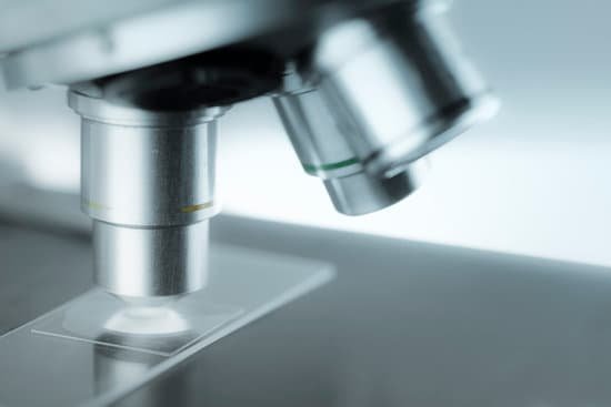
How did the early microscopes work? A Dutch father-son team named Hans and Zacharias Janssen invented the first so-called compound microscope in the late 16th century when they discovered that, if they put a lens at the top and bottom of a tube and looked through it, objects on the other end became magnified.
How did the microscope work? A microscope is an instrument that is used to magnify small objects. … It is through the microscope’s lenses that the image of an object can be magnified and observed in detail. A simple light microscope manipulates how light enters the eye using a convex lens, where both sides of the lens are curved outwards.
What did the first microscopes use? The history of the microscope spans centuries. Roman philosophers mentioned “burning glasses” in their writings but the first primitive microscope was not made until the late 1300’s. Two lenses were placed at opposite ends of a tube. This simple magnifying tube gave birth to the modern microscope.
What were early microscopes used to view? And in the 1670s, he turned his devices to living things—and opened up a new world. He became the first person to observe the inner workings of the body on a microscopic level, seeing bacteria, sperm and even blood cells flowing through capillaries.
How did the early microscopes work? – Related Questions
Will smart phones have microscopes?
DIPLE is far from the only smartphone microscope kit available, and there are lots of ways to achieve similar results. DIY setups are the cheapest, costing as little as $10, but without the same levels of magnification. USB microscopes are another option, ranging in price from $20 to $200 (and up).
What are the main types of microscopes?
There are three basic types of microscopes: optical, charged particle (electron and ion), and scanning probe. Optical microscopes are the ones most familiar to everyone from the high school science lab or the doctor’s office.
Can you see bacteria in water using microscope?
In order to see bacteria, you will need to view them under the magnification of a microscopes as bacteria are too small to be observed by the naked eye. … At high magnification*, the bacterial cells will float in and out of focus, especially if the layer of water between the cover glass and the slide is too thick.
What makes a fluorescent microscope with a uv microscope disadvantages?
Cells are susceptible to phototoxicity, particularly with short-wavelength light. Furthermore, fluorescent molecules have a tendency to generate reactive chemical species when under illumination which enhances the phototoxic effect.
Can aloe vera juice help microscopic colitis?
Aloe vera gel may be beneficial for those with ulcerative colitis. You have to be careful because aloe vera juice, which is more available in stores, has a laxative effect and is therefore a problem for people having diarrhea. Aloe latex (may be called aloe juice) contains strong laxative compounds.
What microscope can be used with live cells?
Microscopy techniques that can capture live cell images include confocal microscopy, phase contrast microscopy, fluorescent microscopy, and quantitative phase contrast microscopy.
What did leeuwenhoek look at through his microscope?
Antonie van Leeuwenhoek used single-lens microscopes, which he made, to make the first observations of bacteria and protozoa. His extensive research on the growth of small animals such as fleas, mussels, and eels helped disprove the theory of spontaneous generation of life.
Do you put used microscope slide into a sharps container?
Glass items contaminated with biohazards, such as pipettes, microscope slides and capillary tubes are also considered “sharps waste.” General Procedures: … biohazardous sharps, the label must be completely covered up.
What is the main function of microscope?
A microscope is an instrument that can be used to observe small objects, even cells. The image of an object is magnified through at least one lens in the microscope. This lens bends light toward the eye and makes an object appear larger than it actually is.
Do compound microscopes use reflected light?
Reflected light is great for seeing surface details in relatively large specimens and is frequently used for low-power microscopy and stereomicroscopic work. … It’s the most frequently used lighting for compound, high-power microscopy. The simplest illuminator is a pivoted mirror to beam external light to the microscope.
Can microscopic hematuria cause anemia?
Despite the sometimes alarming intensity or persistence of hematuria, parents must be informed that, by itself, hematuria rarely causes anemia.
How does saccharomyces microscopically differ from the other specimens?
Saccharomyces is a yeast, a single-celled fungus. How does Saccharomyces microscopically differ from the other specimens? The bacteria were all small, pink clusters of cells with various shapes and sizes. The Saccharomyces was similar in appearance but were blue and slightly larger than the bacteria.
What is ua dipstick w reflex microscopic?
Urinalysis with Reflex to Microscopic – Dipstick urinalysis measures chemical constituents of urine. Microscopic examination helps to detect the presence of cells, bacteria, yeast and other formed elements.
What is microscopic anatomy all about?
Microscopic anatomy (micro; small) is a branch of anatomy that relies on the use of microscopes to examine the smallest structures of the body; tissues, cells, and molecules. … This is known as histology (his-TOL-o-je; the study of tissues).
What is the depth of field microscope quizlet?
The depth of field is a measure of the thickness of a plane of focus. As the magnification increases, the depth of field decreases.
What is the importance of microscope in histology?
LIGHT MICROSCOPES. Light, or optical, microscopes are essential for histological studies because they allow us to visualize cells and morphological features of tissues. Light microscope relies on glass lenses and visible light to magnify tissue samples.
What is the resolving power of the compound light microscope?
The resolving power is the capacity of an instrument to resolve two points that are close together. The resolving power of a compound microscope is 0.25μm.
What is projection microscope?
This microscope is a projection microscope and is the most modern microscope on the tour. As its name suggests, it is a microscope that projects an image of the specimen being examined onto a screen. … Similar to previous compound microscopes on this tour, the light then passes through an objective lens and is magnified.
What are phase rings for on a microscope?
Halos occur in phase contrast microscopy because the circular phase-retarding (and neutral density) ring located in the objective phase plate also transmits a small degree of diffracted light from the specimen (it is not restricted to passing surround waves alone).
Which part of the microscope holds the slide in position?
Stage: The flat platform where you place your slides. Stage clips hold the slides in place. Revolving Nosepiece or Turret: This is the part that holds two or more objective lenses and can be rotated to easily change power.
What are the two parts used to carry the microscope?
The two parts used to carry the microscope was the base and arm carry it both of your hands, so that it won’t slipped off.

