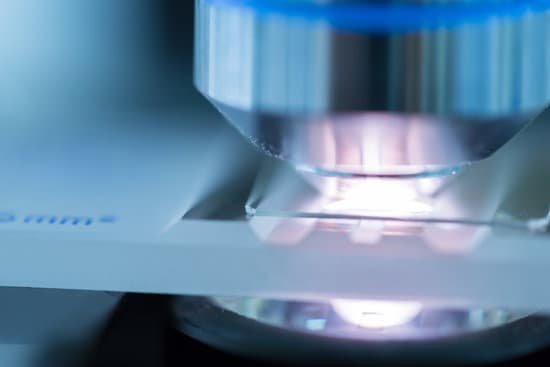
How do objects appear when viewed through a microscope? A microscope is an instrument that can be used to observe small objects, even cells. The image of an object is magnified through at least one lens in the microscope. This lens bends light toward the eye and makes an object appear larger than it actually is.
How are objects viewed with a compound microscope and how do their images appear? The objective lens is positioned close to the object to be viewed. It forms an upside-down and magnified image called a real image because the light rays actually pass through the place where the image lies. The ocular lens, or eyepiece lens, acts as a magnifying glass for this real image.
How does the letter appear while viewing it through the microscope? Notice that it appears upside down when viewed under the microscope. This is a picture of the letter “e” shown at 100X. Notice, that as you increase the power of the lens, your field of view gets smaller.
How can you see something under a microscope? Light from a mirror is reflected up through the specimen, or object to be viewed, into the powerful objective lens, which produces the first magnification. The image produced by the objective lens is then magnified again by the eyepiece lens, which acts as a simple magnifying glass.
How do objects appear when viewed through a microscope? – Related Questions
How do microscopes improve our lives?
A microscope lets the user see the tiniest parts of our world: microbes, small structures within larger objects and even the molecules that are the building blocks of all matter. The ability to see otherwise invisible things enriches our lives on many levels.
How many objective lenses does a compound microscope have?
Objective lens- There is usually 3 or 4 objective lens in the microscope that has different powers. Also, they can magnify objects to a good resolution.
What is the arm on a microscope used for?
Arm connects to the base and supports the microscope head. It is also used to carry the microscope.
Do specimen in a microscope appear upside down?
A microscope is an instrument that magnifies an object. … A specimen that is right-side up and facing right on the microscope slide will appear upside-down and facing left when viewed through a microscope, and vice versa.
What does serratia marcescens look like under a microscope?
Now, Serratia marcescens has a thin peptidoglycan layer, so it doesn’t retain the crystal violet dye during Gram staining. Instead, like any other Gram-negative bacteria, it stains pink with safranin dye. And since it’s a Gram-negative bacillus, it looks like a little pink rod under the microscope.
How microscopes improve our lives?
A microscope lets the user see the tiniest parts of our world: microbes, small structures within larger objects and even the molecules that are the building blocks of all matter. The ability to see otherwise invisible things enriches our lives on many levels.
How did robert hooke observed using his crude microscope?
While observing cork through his microscope, Hooke saw tiny boxlike cavities, which he illustrated and described as cells. He had discovered plant cells! Hooke’s discovery led to the understanding of cells as the smallest units of life—the foundation of cell theory.
How to preserve butterfly wings on microscope slides?
Always keep the wings in alcohol; otherwise they will tear up. After adding a few drops of alcohol and some rubbing with your brush, take a small square of paper towel and soak up the alcohol and scales mix- ture that is on the microscope slide.
How to identify blood cells under microscope?
The identification of blood cells is based primarily on observations of the presence or absence of a nucleus and cytoplasmic granules. Other helpful features are cell size, nuclear size and shape, chromatin appearance, and cytoplasmic staining.
Who created over 500 microscope?
As well as being the father of microbiology, van Leeuwenhoek laid the foundations of plant anatomy and became an expert on animal reproduction. He discovered blood cells and microscopic nematodes, and studied the structure of wood and crystals. He also made over 500 microscopes to view specific objects.
What is a stereo microscope used for?
A stereo microscope is used for low-magnification applications, allowing high-quality, 3D observation of subjects that are normally visible to the naked eye. In life science stereo microscope applications, this could involve the observation of insects or plant life.
How do you kill bugs for microscopic?
“Most of the time in an electron microscope, you have to put mites in a fixative and kill them, and then they tend to shrivel a little,” said Bauchan. “Now, the other way is to freeze them in liquid nitrogen.
How to adjust stereo microscope?
Set the dioptre setting(s) to zero. With the eyes about 10mm away from the eyepieces, adjust the distance between the eyepieces (interpupillary distance) until you see a single image. Adjust the illumination brightness to the desired levels.
Can you look at living microbes in light microscope?
Generally speaking, it is theoretically and practically possible to see living and unstained bacteria with compound light microscopes, including those microscopes which are used for educational purposes in schools.
Who developed the first compound microscope in 1678?
In the late 16th century several Dutch lens makers designed devices that magnified objects, but in 1609 Galileo Galilei perfected the first device known as a microscope. Dutch spectacle makers Zaccharias Janssen and Hans Lipperhey are noted as the first men to develop the concept of the compound microscope.
What does the frame do on a microscope?
The frame consists of the arm, the base and is in essence the bodywork of the microscope. It allows attachment of the focus wheels and the stage to the microscope. A light source used in place of a mirror. Most microscopes do allow manual light adjustment via a wheel located near the base.
What is urinalysis with reflex microscopic?
Urinalysis with Reflex to Microscopic – Dipstick urinalysis measures chemical constituents of urine. Microscopic examination helps to detect the presence of cells, bacteria, yeast and other formed elements.
What is the source of illumination in a microscope?
Incandescent Lamps – Incandescent tungsten-based lamps are the primary illumination source used in modern microscopes, with the exception of those intended for fluorescence microscopy investigations.
What is a microscopic agglutination test?
INDIRECT READING. MAT is a serologic technique that detects agglutinating antibodies against serovar- associated epitopes of the microorganism Leptospira spp. Equal volumes of serial diluted sera are mixed with live leptospires.
How much is a scanning electron microscope?
The price of electron microscopes can also vary by type of electron microscope. The cost of a scanning electron microscope (SEM) can range from $80,000 to $2,000,000. The cost of a transmission electron microscope (TEM) can range from $300,000 to $10,000,000.
How much does an electron microscope magnify?
This makes electron microscopes more powerful than light microscopes. A light microscope can magnify things up to 2000x, but an electron microscope can magnify between 1 and 50 million times depending on which type you use! To see the results, look at the image below.

