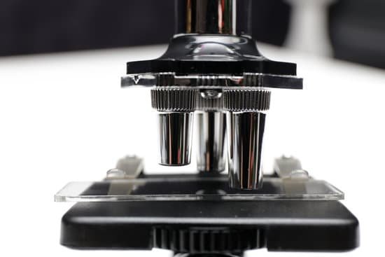
How do you hold a microscope lens? Hold the microscope with one hand around the arm of the device, and the other hand under the base. This is the most secure way to hold and walk with the microscope. Avoid touching the lenses of the microscope. The oil and dirt on your fingers can scratch the glass.
What is the correct way to hold the microscope? Important general rules: Always carry the microscope with 2 hands—place one hand on the microscope arm and the other hand under the microscope base. Do not touch the objective lenses (i.e. the tips of the objectives). Keep the objectives in the scan position and keep the stage low when adding or removing slides.
What holds the objective lenses in place? Stage: The flat platform where you place your slides. Stage clips hold the slides in place. Revolving Nosepiece or Turret: This is the part that holds two or more objective lenses and can be rotated to easily change power. Objective Lenses: Usually you will find 3 or 4 objective lenses on a microscope.
Why is oil used in oil immersion objective? It is best to use an oil-immersed objective at high magnification as the oil compensates for short focal lengths associated with larger magnifications. The oil has a similar refractive value to the glass slides and slipcovers.
How do you hold a microscope lens? – Related Questions
What antidepressants cause microscopic colitis?
Antidepressant agents such as selective serotonin reuptake inhibitors (SSRIs) as a group increase the risk of collagenous colitis, but in this class of medications, sertraline alone significantly raises the risk of lymphocytic colitis (LC).
Where was under the microscope by william j croft published?
Under the microscope: A brief history of microscopy. by William J Croft, Harvard Univ, USA. World Scientific Publishing Pte.
What is the average cost of a light microscope?
Though some high-end light microscopes can cost upwards of $1,000, there are many high-quality options available for far less. You can easily find a decent light microscope for under $100. For a professional-quality light microscope, you can expect to spend $200-$400.
Can lysosomes be resolved with a light microscope?
The limit of resolution of the light microscope is 0.2 µm, while the practical limit of resolution of the electron microscope is about 1 nanometer (nm). Thus, light microscopes allow one to visualize cells and their larger components such as nuclei, nucleoli, secretory granules, lysosomes, and large mitochondria.
Is microscopic hematuria always cancer?
In fact, the overwhelming majority of patients who have microscopic hematuria do not have cancer. Irritation when urinating, urgency, frequency and a constant need to urinate may be symptoms a bladder cancer patient initially experiences.
How to calculate field of view for a microscope?
For instance, if your eyepiece reads 10X/22, and the magnification of your objective lens is 40. First, multiply 10 and 40 to get 400. Then divide 22 by 400 to get a FOV diameter of 0.055 millimeters.
What unit would you use to measure a microscopic organism?
Microbes are broadly defined as organisms that are microscopic. As a result, measuring them can be very difficult. The units used to describe objects on a microscopic length scale are most commonly the Micrometer (oi) – one millionth of 1 meter and smaller units. Most microbes are around 1 micrometer in size.
How to clean a compound light microscope?
To clean microscope eyepiece lenses, breathe condensation onto them and then wipe them with lens tissue. Kim-wipes are made by Kleenex and generally will work well. For stubborn spots, wipe the surface with tissue moistened with 95% alcohol. Wipe the lens dry with a dry tissue.
When was the basic light microscope invented?
In around 1590, Hans and Zacharias Janssen had created a microscope based on lenses in a tube [1]. No observations from these microscopes were published and it was not until Robert Hooke and Antonj van Leeuwenhoek that the microscope, as a scientific instrument, was born.
What is the resolving power of a microscope?
The resolving power of a microscope is taken as the ability to distinguish between two closely spaced Airy disks (or, in other words, the ability of the microscope to distinctly reveal adjacent structural detail). It is this effect of diffraction that limits a microscope’s ability to resolve fine details.
What is a scanning electron microscope work?
A scanning electron microscope (SEM) scans a focused electron beam over a surface to create an image. The electrons in the beam interact with the sample, producing various signals that can be used to obtain information about the surface topography and composition.
Where is the coarse focus knob on a microscope?
Coarse Adjustment Knob- The coarse adjustment knob located on the arm of the microscope moves the stage up and down to bring the specimen into focus. The gearing mechanism of the adjustment produces a large vertical movement of the stage with only a partial revolution of the knob.
What are the 2 types of lenses on a microscope?
The common light microscope used in the laboratory is called a compound microscope because it contains two types of lenses that function to magnify an object. The lens closest to the eye is called the ocular, while the lens closest to the object is called the objective.
Why is it unclear who invented the compound microscope?
The development of the microscope allowed scientists to make new insights into the body and disease. It’s not clear who invented the first microscope, but the Dutch spectacle maker Zacharias Janssen (b. 1585) is credited with making one of the earliest compound microscopes (ones that used two lenses) around 1600.
Do objects in a microscope appear upside down?
A microscope is an instrument that magnifies an object. … The optics of a microscope’s lenses change the orientation of the image that the user sees. A specimen that is right-side up and facing right on the microscope slide will appear upside-down and facing left when viewed through a microscope, and vice versa.
Why are electron microscopes so expensive?
Cost – Electron microscopes are expensive pieces of highly specialized equipment. … Also, as electron microscopes are highly sensitive, magnetic fields and vibrations caused by other lab equipment may interfere with their operation.
What is an ocular in a microscope?
The eyepiece, or ocular lens, is the part of the microscope that magnifies the image produced by the microscope’s objective so that it can be seen by the human eye.
How to pick up a microscope?
Hold the microscope with one hand around the arm of the device, and the other hand under the base. This is the most secure way to hold and walk with the microscope. Avoid touching the lenses of the microscope. The oil and dirt on your fingers can scratch the glass.
Who invented the first microscope in 1590?
Lens Crafters Circa 1590: Invention of the Microscope. Every major field of science has benefited from the use of some form of microscope, an invention that dates back to the late 16th century and a modest Dutch eyeglass maker named Zacharias Janssen.
What is other microscopic hematuria?
“Microscopic” means something is so small that it can only be seen through a special tool called a microscope. “Hematuria” means blood in the urine. So, if you have microscopic hematuria, you have red blood cells in your urine. These blood cells are so small, though, you can’t see the blood when you urinate.

