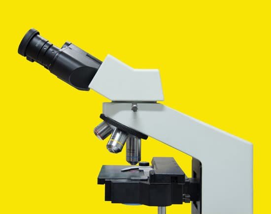
How do you improve contrast on a microscope? Contrast may be improved by placing suitable apertures or filters within the optical path, either in the illuminating system alone (dark ground or Rheinberg illumination), or in conjugate planes in the imaging system (e.g. for phase contrast, differential interference contrast or polarised light microscopy).
How can contrast be improved? Contrast can also be increased by physical modification of the microscope optical components and illumination mode, as well as manipulation of the final image through photographic or digital electronic techniques.
What methods are used to improve contrast in bright field microscopy? Polarized light microscopy is a contrast-enhancing technique that dramatically improves the quality of an image acquired with birefringent materials when compared to other techniques such as brightfield and darkfield illumination, phase contrast, differential interference contrast, fluorescence, and Hoffman modulation …
What component of the microscope is used to enhance contrast? Iris Diaphragm: Found on high power microscopes under the stage, the diaphragm is, typically, a five hole-disc with each hole having a different diameter. It is used to vary the light that passes through the stage opening and helps to adjust both the contrast and resolution of a specimen.
How do you improve contrast on a microscope? – Related Questions
What do ribosomes look like under a microscope?
Ribosomes – which cannot be seen with a light microscope, but only using an electron microscope – are the site of protein synthesis. Even at very high magnification, they look like (pairs of) dots.
Why is the light microscope useful?
Light microscopes are an invaluable analytical tool that has the potential to allow scientific investigators to view objects at 1000 times their original size. Light microscopes magnify and resolve the image of an object that is otherwise invisible to naked eye.
What is meant by the total magnification of a microscope?
Magnification: the process of enlarging the size of an object, as an optical image. Total magnification: In a compound microscope the total magnification is the product of the objective and ocular lenses (see figure below). … Thus, this increases the image’s contrast between the specimen and the medium.
What is the definition of transmission electron microscope in biology?
Transmission electron microscopes (TEM) are microscopes that use a particle beam of electrons to visualize specimens and generate a highly-magnified image. TEMs can magnify objects up to 2 million times. In order to get a better idea of just how small that is, think of how small a cell is.
How to tell if bacteria is alive under a microscope?
Re: How can I tell if my microorganisms are alive or not? The best way to find out if the amoeba and paramecium are alive is to look at them under the microscope and see them moving. The E coli and Bacillus you can spread on nutrient agar in a Petri dish and see if they form colonies.
What type of microscope has the largest magnification?
Out of all types of microscopes, the electron microscope has the greatest capability in achieving high magnification and resolution levels, enabling us to look at things right down to each individual atom.
How much electron microscope cost?
The price of a new electron microscope can range from $80,000 to $10,000,000 depending on certain configurations, customizations, components, and resolution, but the average cost of an electron microscope is $294,000. The price of electron microscopes can also vary by type of electron microscope.
What can you do with electron microscopes?
Electron microscopes are used to investigate the ultrastructure of a wide range of biological and inorganic specimens including microorganisms, cells, large molecules, biopsy samples, metals, and crystals. Industrially, electron microscopes are often used for quality control and failure analysis.
What is the measure of clarity on a microscope?
The resolution of a microscope or lens is the smallest distance by which two points can be separated and still be distinguished as separate objects. The smaller this value, the higher the resolving power of the microscope and the better the clarity and detail of the image.
What do electron microscopes do?
The electron microscope uses a beam of electrons and their wave-like characteristics to magnify an object’s image, unlike the optical microscope that uses visible light to magnify images. … This beam is focused onto the sample using a magnetic lens.
Where is the slide on a microscope?
The bottom layer is the slide. Next is the liquid sample. A small square of clear glass or plastic (a coverslip) is placed on top of the liquid to minimize evaporation and protect the microscope lens from exposure to the sample.
What is microscopic disc surgery?
Microdiscectomy, also sometimes called microdecompression or microdiskectomy, is a minimally invasive surgical procedure performed on patients with a herniated lumbar disc. During this surgery, a surgeon will remove portions of the herniated disc to relieve pressure on the spinal nerve column.
Do confocal microscopes take pictures?
As discussed above, the confocal fluorescence microscope consists of multiple laser excitation sources, a scan head with optical and electronic components, electronic detectors (usually photomultipliers), and a computer for acquisition, processing, analysis, and display of images.
Why do scientists use microscopes in their work?
Scientists use microscopes to observe objects too small to view with the human eye. Microscopes can magnify an image hundreds of times while…
When did the first microscope come out?
The first compound microscopes date to 1590, but it was the Dutch Antony Van Leeuwenhoek in the mid-seventeenth century who first used them to make discoveries. When the microscope was first invented, it was a novelty item.
Can dna double helix be seen with a microscope?
Under a microscope, the familiar double-helix molecule of DNA can be seen. Because it is so thin, DNA cannot be seen by the naked eye unless its strands are released from the nuclei of the cells and allowed to clump together.
What is the specimen preparation for a light microscope?
There are two basic types of preparation used to view specimens with a light microscope: wet mounts and fixed specimens. The simplest type of preparation is the wet mount, in which the specimen is placed on the slide in a drop of liquid.
How should you always carry a microscope?
Always keep your microscope covered when not in use. Always carry a microscope with both hands. Grasp the arm with one hand and place the other hand under the base for support.
When did the microscope invented?
Lens Crafters Circa 1590: Invention of the Microscope. Every major field of science has benefited from the use of some form of microscope, an invention that dates back to the late 16th century and a modest Dutch eyeglass maker named Zacharias Janssen.

