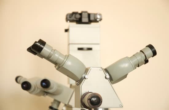
How does a compound microscope work? A compound microscope uses two or more lenses to produce a magnified image of an object, known as a specimen, placed on a slide (a piece of glass) at the base. … By raising and lowering the stage, you move the lenses closer to or further away from the object you’re examining, adjusting the focus of the image you see.
How does a compound microscope work step by step? Turn the revolving turret (2) so that the lowest power objective lens (eg. 4x) is clicked into position. Place the microscope slide on the stage (6) and fasten it with the stage clips. Look at the objective lens (3) and the stage from the side and turn the focus knob (4) so the stage moves upward.
How does a compound lens work? A compound lens uses multiple lenses. The most obvious example of a simple lens is a magnifying glass, which uses a single lens to magnify an object, while an example of a compound lens is a compound microscope, which uses multiple lenses to increase the viewer’s capacity to magnify an object.
How does a compound microscope form an image? The classic compound microscope magnifies in two steps: first with an objective lens that produces an enlarged image of the object in a ‘real’ image plane. This real image is then magnified by the ocular lens or eyepiece to produce the virtual image. Two convex lenses can form a microscope.
How does a compound microscope work? – Related Questions
What is the ocular magnification for scanning on a microscope?
A scanning objective lens provides the lowest magnification power of all objective lenses. 4x is a common magnification for scanning objectives and, when combined with the magnification power of a 10x eyepiece lens, a 4x scanning objective lens gives a total magnification of 40x.
How to use a usb microscope?
Plug the device into any open USB port on the computer or the television. Hold the microscope and lightly touch the lens to the specimen. The image should now be visible on the monitor or television screen. These microscopes should only be used to examine dry specimens.
Is a magnifying glass the same as a microscope?
Compound Microscope. A simple microscope uses a single lens, so magnifying glasses are simple microscopes. … Stereoscopic microscopes use two oculars or eyepieces, one for each eye, to allow binocular vision and provide a three-dimensional view of the object.
How much is a light microscope?
Though some high-end light microscopes can cost upwards of $1,000, there are many high-quality options available for far less. You can easily find a decent light microscope for under $100. For a professional-quality light microscope, you can expect to spend $200-$400.
How to focus compound microscope?
Look at the objective lens (3) and the stage from the side and turn the focus knob (4) so the stage moves upward. Move it up as far as it will go without letting the objective touch the coverslip. Look through the eyepiece (1) and move the focus knob until the image comes into focus.
What does the objective lens do on a microscope do?
The objective, located closest to the object, relays a real image of the object to the eyepiece. This part of the microscope is needed to produce the base magnification. The eyepiece, located closest to the eye or sensor, projects and magnifies this real image and yields a virtual image of the object.
What did robert koch discovered with a microscope?
It was there that he began studying the biology of bacteria. At the time there were still no electron microscopes and bacteria were the smallest pathogens that could be seen with a standard microscope. Koch discovered the anthrax bacterium, and was the first to describe how it was transmitted.
Why are scanning electron microscope images black and white?
Why do electron microscopes produce black and white images? The reason is pretty basic: color is a property of light (i.e., photons), and since electron microscopes use an electron beam to image a specimen, there’s no color information recorded.
What is the preferred microscope for counting manual platelet counts?
The determination is done by placing a small volume of diluted whole blood that was treated with a red cell lysing reagent, such as ammonium oxalate, in a counting chamber (hemocytometer), and counting platelets using phase contrast light microscopy.
What types of electron microscopes are there?
There are two main types of electron microscope – the transmission EM (TEM) and the scanning EM (SEM). The transmission electron microscope is used to view thin specimens (tissue sections, molecules, etc) through which electrons can pass generating a projection image.
How are microscopes made?
Lenses are given an antireflective coating, usually of magnesium fluoride. The eyepiece, the objective, and most of the hardware components are made of steel or steel and zinc alloys. A child’s microscope may have an external body shell made of plastic, but most microscopes have an body shell made of steel.
What are the limitations of a compound light microscope?
The magnifying power of a compound light microscope is limited to 2000 times. Certain specimens, such as viruses, atoms, and molecules can’t be viewed with it.
Can white blood cells be seen under a light microscope?
Given that all white blood cells are over 5 micrometers in diameter, they are large enough to be seen using a typical optical microscope (compound microscope).
How to read the objective lens of microscope?
Microscope objective lenses will often have four numbers engraved on the barrel in a 2×2 array. The upper left number is the magnification factor of the objective. For example, 4x, 10x, 40x, and 100x. The upper right number is the numerical aperture of the objective.
What is appearance fluid microscopic exam?
Microscopic examination – a laboratory professional places a sample of your fluid on a slide and examines it using a microscope, counting any white blood cells (WBCs) and red blood cells (RBCs) and looking for bacteria or fungi.
When microscopes are parfocal?
To simplify, if a compound microscope is parfocal, it means that when you change magnification sequentially (ex. 4x to 10x to 40x to 100x), it will only require a very slight turn of the fine focus knob with each increase or decrease to get the image in focus.
What size can you see with transmission electron microscopes?
They are also the most powerful microscopic tool available to-date, capable of producing high-resolution, detailed images 1 nanometer in size.
What does the microscope slide rests on?
Stage: The platform the slide rests on while being viewed. The stage has a hole in it to permit light to pass through both it and the specimen. The mechanical stage permits precise movement of the specimen.
What microscope has the largest magnification?
Out of all types of microscopes, the electron microscope has the greatest capability in achieving high magnification and resolution levels, enabling us to look at things right down to each individual atom.
What is light stereo microscope?
The stereo, stereoscopic or dissecting microscope is an optical microscope variant designed for low magnification observation of a sample, typically using light reflected from the surface of an object rather than transmitted through it.
What is the importance of microscope in science?
The invention of the microscope allowed scientists to see cells, bacteria, and many other structures that are too small to be seen with the unaided eye. It gave them a direct view into the unseen world of the extremely tiny. You can get a glimpse of that world in Figure below.

