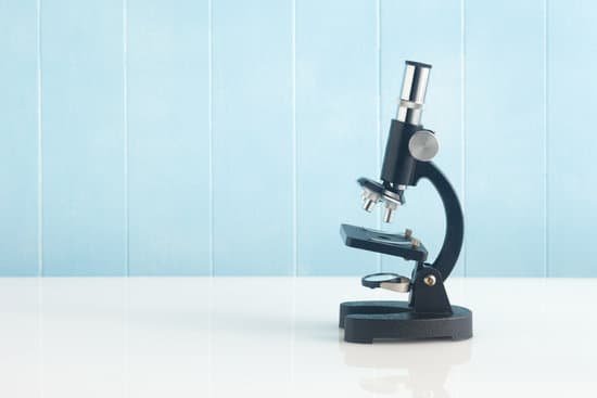
How does a microscope help scientists? A microscope is an instrument that is used to magnify small objects. Some microscopes can even be used to observe an object at the cellular level, allowing scientists to see the shape of a cell, its nucleus, mitochondria, and other organelles.
How do microscopes most help scientists? Microscopes help the scientists to study the microorganisms, the cells, the crystalline structures, and the molecular structures, They are one of the most important diagnostic tools when the doctors examine the tissue samples.
How did the microscope improve science? Despite some early observations of bacteria and cells, the microscope impacted other sciences, notably botany and zoology, more than medicine. Important technical improvements in the 1830s and later corrected poor optics, transforming the microscope into a powerful instrument for seeing disease-causing micro-organisms.
What have microscopes helped us discover? Microscopes allow humans to see cells that are too tiny to see with the naked eye. Therefore, once they were invented, a whole new microscopic world emerged for people to discover. On a microscopic level, new life forms were discovered and the germ theory of disease was born.
How does a microscope help scientists? – Related Questions
What are the limits of a compound microscope?
The magnifying power of a compound light microscope is limited to 2000 times. Certain specimens, such as viruses, atoms, and molecules can’t be viewed with it.
Why look at urine under a microscope?
This test looks at a sample of your urine under a microscope. It can see cells from your urinary tract, blood cells, crystals, bacteria, parasites, and cells from tumors. This test is often used to confirm the findings of other tests or add information to a diagnosis.
Can a usb microscope be used with android phone?
AbleScope is a special APP to our customers who want to connect the USB Microscope and USB Endoscope to the Android Smart Phones and Tablet without rooting. AbleScope is mainly developed for Android devices that carrying Qualcomm chips as the CPU in the smart phones and tablet.
What is rheostat on a microscope?
rheostat – alters the current applied to the lamp to control the intensity of the light produced condenser – lens system that aligns and focuses the light from the lamp onto the specimen diaphragms or pinhole apertures – placed in the light path to alter the amount of light that reaches the condenser (for enhancing …
Why do you put a coverslip on a microscope slide?
When viewing any slide with a microscope, a small square or circle of thin glass called a coverslip is placed over the specimen. It protects the microscope and prevents the slide from drying out when it’s being examined.
How to see giardia under microscope?
In bright-field microscopy, cysts appear ovoid to ellipsoid in shape and usually measure 11 to 14 µm (range: 8 to 19 µm). Immature and mature cysts have 2 and 4 nuclei, respectively. Intracytoplasmic fi brils are visible in cysts.
How does a light microscope work for kids?
microscopes, also called light microscopes, work like magnifying glasses. They use lenses, which are curved pieces of glass or plastic that bend light. The object to be studied sits under a lens. As light passes from the object through the lens, the lens makes the object look bigger.
What type of image does a scanning electron microscope produce?
A scanning electron microscope (SEM) is a type of microscope which uses a focused beam of electrons to scan a surface of a sample to create a high resolution image. SEM produces images that can show information on a material’s surface composition and topography.
What is the function of the arm of a microscope?
Arm connects to the base and supports the microscope head. It is also used to carry the microscope.
Does an electron microscope have a radioactive sources?
The radiation safety concerns are related to the electrons that are backscattered from the sample, as well as X-rays produced in the process. … However, scanning electron microscopes are radiation-generating devices and should be at least inventoried.
How to select a microscope?
When Choosing the most important lens in a microscope is the one closest to the specimen. Compound microscopes generally have three, four or five objective lenses, so you can select different magnification levels. The higher the number, or power, of an objective lens, the finer the detail.
What cells cannot be seen with a light microscope?
Some cell parts, including ribosomes, the endoplasmic reticulum, lysosomes, centrioles, and Golgi bodies, cannot be seen with light microscopes because these microscopes cannot achieve a magnification high enough to see these relatively tiny organelles.
Who invented microscope netherlands?
Dutch scientist Antoine van Leeuwenhoek designed high-powered single lens microscopes in the 1670s. With these he was the first to describe sperm (or spermatozoa) from dogs and humans. He also studied yeast, red blood cells, bacteria from the mouth and protozoa.
What does each part of a light microscope do?
Lenses – form the image objective lens – gathers light from the specimen eyepiece – transmits and magnifies the image from the objective lens to your eye nosepiece – rotating mount that holds many objective lenses tube – holds the eyepiece at the proper distance from the objective lens and blocks out stray light.
How is your specimen different when viewed under the microscope?
The optics of a microscope’s lenses change the orientation of the image that the user sees. A specimen that is right-side up and facing right on the microscope slide will appear upside-down and facing left when viewed through a microscope, and vice versa.
Why is a microscope needed to study cells?
A cell is the smallest unit of life. Most cells are so small that they cannot be viewed with the naked eye. Therefore, scientists must use microscopes to study cells. Electron microscopes provide higher magnification, higher resolution, and more detail than light microscopes.
How to collect pond water for microscope?
A good method of collecting them is to lower a jar upside down until it is positioned just above the mud surface. Then slowly let the air escape so the top layer will be sucked into the jar. You can move the jar slowly when tilting so you collect from a larger area.
What do the objective lenses do on a microscope?
The objective, located closest to the object, relays a real image of the object to the eyepiece. This part of the microscope is needed to produce the base magnification. The eyepiece, located closest to the eye or sensor, projects and magnifies this real image and yields a virtual image of the object.
Which types of microscope gives the highest amount of magnification?
Out of all types of microscopes, the electron microscope has the greatest capability in achieving high magnification and resolution levels, enabling us to look at things right down to each individual atom.
How did microscopes improve our understanding of living things?
Microscopes allow humans to see cells that are too tiny to see with the naked eye. Therefore, once they were invented, a whole new microscopic world emerged for people to discover. On a microscopic level, new life forms were discovered and the germ theory of disease was born.
What is the primary microscopic origin of the magnetic field?
The origin of the magnetic moments responsible for magnetization can be either microscopic electric currents resulting from the motion of electrons in atoms, or the spin of the electrons or the nuclei.

