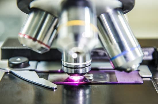
How does a water drop microscope work? The surface of a water drop curves outward to make a dome. This outward, or convex, curvature bends light rays inward. The result is an enlarged image on the retina of your eye.
How does a water microscope work? The water forms a convex lens shape which helps to magnify your specimen. … Convex lens: This kind of lens bends or protrudes outwards so it looks fatter or domed on each side, as light passes through this lens it converges (comes to a point) which makes things look bigger.
How do you make a water drop microscope? The rays of light converge when they pass through the drops of water. Thus, the drop of water behaves like a convex lens.
Are water droplets concave or convex? The magnification is defined as the image size divided by the object size, so this was 1554/150, or approximately 10X. The diameter of the water lens for this measurement wasn’t recorded.
How does a water drop microscope work? – Related Questions
Do microscopes used to test blood?
For a blood smear test, a laboratory professional examines the slide under a microscope and looks at the size, shape, and number of different types of blood cells.
What are the objectives on a compound microscope?
A compound microscope has multiple lenses: the objective lens (typically 4x, 10x, 40x or 100x) is compounded (multiplied) by the eyepiece lens (typically 10x) to obtain a high magnification of 40x, 100x, 400x and 1000x. Higher magnification is achieved by using two lenses rather than just a single magnifying lens.
Are bed bugs microscopic or bigger?
Bedbugs are reddish brown insects that are small in size, but not too small to see with the naked eye. They’re usually about 3⁄16-inches long and have six legs.
Can i buy a camera for my microscope?
Microscope USB cameras include software that allow you to view a live image from the microscope on the computer. … USB microscope cameras are available for high resolution, high speed (fast frame rates), fluorescence work, and extended depth of focus.
Do the microscope samples in sims 4 do anything?
Microscope Prints are great to decorate your house with just like the Space Prints Collection. They have a lot of different strong colors that will go great with most of the standard interior colors. The prints come in different sizes up to 3 tiles big!
When was the first light microscope invented and by who?
In 1609, Galileo Galilei made a microscope by converting one of his telescopes. It had a diverging lens as an eyepiece and a converging lens as an objective. An early microscope made of two converging lenses was presented around 1620 by the astronomer Cornelius Drebbel.
When was the microscope invented wikipedia?
13th century — The increase in use of lenses in eyeglasses probably led to the wide spread use of simple microscopes (single lens magnifying glasses) with limited magnification. 1590 — earliest date of a claimed Hans Martens/Zacharias Janssen invention of the compound microscope (claim made in 1655).
How to use a pocket microscope?
Place the microscope directly on the top of the object of observation and press the LED On/Off Button. 2. Slide the Zoom Lever to adjust the desired degree of magnification (from 20x to 40x) 3. Turn the Focusing wheel gradually until the image is clear and sharp.
Can microscopic colitis cause bleeding?
Pus and fluid also are secreted into the colon and add to the diarrhea. The redness, bleeding of the lining with gentle rubbing (friability), and ulcerations in the lining of the colon contribute to rectal bleeding.
How do microscopic invertebrates differ from protozoa?
Protozoa are single celled organisms that are very diverse groups. Invertebrates are multi-cellular animals without a backbone or bony skeleton. …
How to describe cell appearance under the microscope?
Under a low power microscope, the cell membrane is observed as a thin line, while the cytoplasm is completely stained. The cell organelles are seen as tiny dots throughout the cytoplasm, whereas the nucleus is seen as a thick drop.
How does an image appear under a microscope?
A microscope is an instrument that can be used to observe small objects, even cells. The image of an object is magnified through at least one lens in the microscope. This lens bends light toward the eye and makes an object appear larger than it actually is.
What are two types of light microscopes?
used to view specimens are both simple and compound light microscopes, all using lenses. The difference is simple light microscopes use a single lens for magnification while compound lenses use two or more lenses for magnifications.
Do condoms have holes microscopic holes?
Natural “skin” condoms (made of lamb membrane) protect against pregnancy but contain pores (microscopic holes) that are large enough to let viruses pass through (but not large enough to let sperm pass through).
Can an mri detect microscopic scar tissue?
Magnetic resonance imaging (MRI) is a powerful tool that can detect microscopic injuries including damage to nerve fibers. MRI can also reveal very small bleeds (microhemorrhage), small bruises, and scarring (gliosis), that are not visible on a CT scan.
How do you clean your microscope slides?
When slides get soiled, you can clean them with soapy water or isopropyl alcohol. Do not immerse slides in water or soak them in it. This loosens the cover glass adhesive, causing the cover glass to come off and possibly ruin the slide.
What is microscopic anatomy in biology?
Microscopic anatomy: The study of normal structure of an organism under the microscope. Known among medical students simply as ‘micro.
What is microscope objective working distance?
Microscope objectives are generally designed with a short free working distance, which is defined as the distance from the front lens element of the objective to the closest surface of the coverslip when the specimen is in sharp focus.
Did hooke’s microscope use light?
For illumination purposes, Hooke designed an ingenious method of concentrating light on his specimens. He passed light generated from an oil lamp through a water-filled glass flask to diffuse the light and provide better illumination for the samples.
Can you see an amoeba without a microscope?
Most Amoeba are microscopic and cannot be seen without a microscope. However, some types of Amoeba are large enough to be seen with the unaided eye….
Can dna only be seen through an electron microscope?
To view the DNA as well as a variety of other protein molecules, an electron microscope is used. Whereas the typical light microscope is only limited to a resolution of about 0.25um, the electron microscope is capable of resolutions of about 0.2 nanometers, which makes it possible to view smaller molecules.

