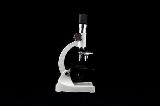
How does scabies look under microscope? Most people with scabies only carry 10 to 15 mites at any given time, and each mite is less than half a millimeter long. This makes them very difficult to spot. To the naked eye, they may look like tiny black dots on the skin. A microscope can identify mites, eggs, or fecal matter from a skin scraping.
Can you see scabies with a magnifying glass? Scabies mites can be seen with a magnifying glass or microscope. The scabies mites crawl but are unable to fly or jump. They are immobile at temperatures below 20º C, although they may survive for prolonged periods at these temperatures. Scabies infestation occurs worldwide and is very common.
Can you see scabies with the human eye? Scabies is caused by the mite known as the Sarcoptes scabiei. These mites are so tiny that they can’t be seen by the human eye. When viewed by a microscope, you’d see they have a round body and eight legs.
What kind of microscope can see scabies? Dermoscopy is an alternative technique for diagnosing scabies. An illuminated magnifier, also known as an epiluminescent stereomicroscope (magnification × 20–60) is required. The hand-held device, designed in 1996 by the dermatologist JF Kreusch, is held perpendicular to the skin (Figure 1).
How does scabies look under microscope? – Related Questions
Do electron microscopes look at living cells?
Electron microscopes are the most powerful type of microscope, capable of distinguishing even individual atoms. However, these microscopes cannot be used to image living cells because the electrons destroy the samples.
What is meant by depth of field in a microscope?
Also known as ‘depth of field’, this is the distance (measured in the. direction of the optical axis) between the two planes which define the. limits of acceptable image sharpness when the microscope is focused. on an object.
How to calculate cell size microscope equation?
L, where D is the diameter of the viewing field, E is the estimated number of cells, and L is the length of the cell.
Who was the first person to invent the microscope?
A Dutch father-son team named Hans and Zacharias Janssen invented the first so-called compound microscope in the late 16th century when they discovered that, if they put a lens at the top and bottom of a tube and looked through it, objects on the other end became magnified.
What can scanning electron microscopes see?
Because of its great depth of focus, a scanning electron microscope is the EM analog of a stereo light microscope. It provides detailed images of the surfaces of cells and whole organisms that are not possible by TEM. It can also be used for particle counting and size determination, and for process control.
What is the high power objective on a microscope?
The high-powered objective lens (also called “high dry” lens) is ideal for observing fine details within a specimen sample. The total magnification of a high-power objective lens combined with a 10x eyepiece is equal to 400x magnification, giving you a very detailed picture of the specimen in your slide.
How to see sperm under a microscope?
You can view sperm at 400x magnification. You do NOT want a microscope that advertises anything above 1000x, it is just empty magnification and is unnecessary. In order to examine semen with the microscope you will need depression slides, cover slips, and a biological microscope.
What could microscopic blood in urine mean?
Microscopic urinary bleeding is a common symptom of glomerulonephritis, an inflammation of the kidneys’ filtering system. Glomerulonephritis may be part of a systemic disease, such as diabetes, or it can occur on its own.
Who made the microscope in 1665?
Interested in learning more about the microscopic world, scientist Robert Hooke improved the design of the existing compound microscope in 1665. His microscope used three lenses and a stage light, which illuminated and enlarged the specimens.
When are upright microscopes used?
In cell biology, upright microscopes are used for phase contrast or widefield fluorescence microscopy of living cells or samples that are squeezed between a slide and coverslip. An additional application is the microscopy of fixed cells or tissue sections.
How have electron microscopes helped our understanding of cells?
The development of the electron microscopes therefore helped scientists to learn about the sub-cellular structures involved in aerobic respiration called mitochondria . The scientists developed their explanations about how the structure of the mitochondria allowed it to efficiently carry out aerobic respiration.
How does the ion microscope function?
The ions are imaged onto a screen, reflecting the crystal structure of the surface sites in which the gas had been adsorbed. The intense field in created by applying a high potential to a very sharp needle, otherwise known as a field emission tip, which has a radius of curvature ∼50 nm.
Can kidney stones cause microscopic blood in urine?
The stones are generally painless, so you probably won’t know you have them unless they cause a blockage or are being passed. Then there’s usually no mistaking the symptoms — kidney stones, especially, can cause excruciating pain. Bladder or kidney stones can also cause both gross and microscopic bleeding.
What type of machine is a microscope?
Optical. The most common type of microscope (and the first invented) is the optical microscope. This is an optical instrument containing one or more lenses producing an enlarged image of a sample placed in the focal plane.
Does sem microscope use fluorescence?
Fluorescence techniques are widely used in biological research to examine molecular localization, while electron microscopy can provide unique ultrastructural information. … We successfully demonstrated that the FL-SEM is a simple and practical tool for correlative fluorescence and electron microscopy.
Does shaving cause microscopic tears?
According to a 2017 study , about 25% of people injure themselves while grooming their pubic hair. Individuals with skin conditions may be more prone to cuts and wounds during hair removal. In addition to larger cuts or tears, all forms of hair removal can cause microscopic wounds.
How does magnification occur in an electron microscope?
The electron microscope uses a beam of electrons and their wave-like characteristics to magnify an object’s image, unlike the optical microscope that uses visible light to magnify images. … This beam is focused onto the sample using a magnetic lens.
Which of the following is also known as microscopic anatomy?
Histology, also known as microscopic anatomy or microanatomy, is the branch of biology which studies the microscopic anatomy of biological tissues.
What does cartilage look like under a microscope?
It contains no nerves or blood vessels, and its structure is relatively simple. If a thin slice of cartilage is examined under the microscope, it will be found to consist of cells of a rounded or bluntly angular form, lying in groups of two or more in a granular or almost homogeneous matrix.
What should sperm look like under a microscope?
The air-fixed, stained spermatozoa are observed under a bright-light microscope at 400x or 1000x magnification. Their viability and mor- phology can be analysed at the same time. Those appearing red-pink in colour have a damaged membrane whereas white sperm are viable, as in Photo 2.
What did robert brown see in his microscope?
In 1827, while examining grains of pollen of the plant Clarkia pulchella suspended in water under a microscope, Brown observed minute particles, now known to be amyloplasts (starch organelles) and spherosomes (lipid organelles), ejected from the pollen grains, executing a continuous jittery motion.

