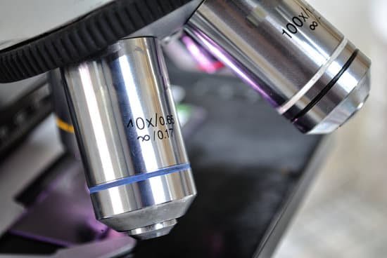
How good a microscope do you use to see cells? Most cells are so small that they cannot be viewed with the naked eye. Therefore, scientists must use microscopes to study cells. Electron microscopes provide higher magnification, higher resolution, and more detail than light microscopes.
How strong of a microscope Do you need to see cells? Magnification of 400x is the minimum needed for studying cells and cell structure.
What type of microscope is best to see cells? Compound microscopes are light illuminated. The image seen with this type of microscope is two dimensional. This microscope is the most commonly used. You can view individual cells, even living ones.
Can a microscope see cells? A microscope is an instrument that can be used to observe small objects, even cells. The image of an object is magnified through at least one lens in the microscope.
How good a microscope do you use to see cells? – Related Questions
What an electron microscope is used for?
Electron microscopy (EM) is a technique for obtaining high resolution images of biological and non-biological specimens. It is used in biomedical research to investigate the detailed structure of tissues, cells, organelles and macromolecular complexes.
How do you use a compound light microscope?
Turn the revolving turret (2) so that the lowest power objective lens (eg. 4x) is clicked into position. Place the microscope slide on the stage (6) and fasten it with the stage clips. Look at the objective lens (3) and the stage from the side and turn the focus knob (4) so the stage moves upward.
What is scanning power on a microscope?
A scanning objective lens provides the lowest magnification power of all objective lenses. 4x is a common magnification for scanning objectives and, when combined with the magnification power of a 10x eyepiece lens, a 4x scanning objective lens gives a total magnification of 40x.
Which microscope does not invert the image?
Quite a few microscopes, including electron microscopes and digital microscopes, will not show you inverted images. Binocular and dissecting microscopes will also not show an inverted image because of their increased level of magnification.
Does a microscope have an inverted final image?
The purpose of a microscope is to create magnified images of small objects, and both lenses contribute to the final magnification. … The image produced by the eyepiece is a magnified virtual image. The final image remains inverted but is farther from the observer than the object, making it easy to view.
How to measure the magnification of a microscope?
To calculate the total magnification of the compound light microscope multiply the magnification power of the ocular lens by the power of the objective lens. For instance, a 10x ocular and a 40x objective would have a 400x total magnification. The highest total magnification for a compound light microscope is 1000x.
What is a microscope slide in chemistry?
A microscope slide is a thin flat piece of glass, typically 75 by 26 mm (3 by 1 inches) and about 1 mm thick, used to hold objects for examination under a microscope. Typically the object is mounted (secured) on the slide, and then both are inserted together in the microscope for viewing.
Why would a biology use microscope?
The microscope is important because biology mainly deals with the study of cells (and their contents), genes, and all organisms. Some organisms are so small that they can only be seen by using magnifications of ×2000−×25000 , which can only be achieved by a microscope. Cells are too small to be seen with the naked eye.
Why is microscopic examination of urine sediment helpful in diagnosis?
This test performs very favorably as a urinary “biomarker” for a number of acute kidney diseases. When used properly, urine sediment findings alert health care providers to the presence of kidney disease, while also providing diagnostic information that often identifies the compartment of kidney injury.
What compound light microscope used for?
Typically, a compound microscope is used for viewing samples at high magnification (40 – 1000x), which is achieved by the combined effect of two sets of lenses: the ocular lens (in the eyepiece) and the objective lenses (close to the sample).
What are cells you can see without a microscope?
Other organisms, however, have only one cell in their entire body, and humans can see some of these single-cell organisms with the naked eye. Human egg cells, unusually large bacteria, some amoebas and squid nerve cells make up this list.
How is a compound microscope different from a magnifying glass?
One difference between a magnifying glass and a compound light microscope is that a magnifying glass uses one lens to magnify an object while a compound microscope uses two or more lenses.
What is a numerical aperture microscope?
Numerical Aperture and Resolution. The numerical aperture of a microscope objective is the measure of its ability to gather light and to resolve fine specimen detail while working at a fixed object (or specimen) distance. … The smaller the object, the more pronounced the diffraction of incident light rays will be.
Why are the stage and diaphragm important parts of microscope?
Aperture is the hole in the stage through which the base (transmitted) light reaches the stage. … Iris Diaphragm controls the amount of light reaching the specimen. It is located above the condenser and below the stage. Most high quality microscopes include an Abbe condenser with an iris diaphragm.
What is the prefix of the word microscope?
An easy way to remember that the prefix micro- means “small” is through the word microscope, an instrument which allows the viewer to see “small” living things.
What is the use of objective lens on a microscope?
The objective, located closest to the object, relays a real image of the object to the eyepiece. This part of the microscope is needed to produce the base magnification. The eyepiece, located closest to the eye or sensor, projects and magnifies this real image and yields a virtual image of the object.
How does a dissecting microscope work?
The stereo, stereoscopic or dissecting microscope is an optical microscope variant designed for low magnification observation of a sample, typically using light reflected from the surface of an object rather than transmitted through it.
Why do electron microscopes have a higher resolution?
Electron microscopes differ from light microscopes in that they produce an image of a specimen by using a beam of electrons rather than a beam of light. Electrons have much a shorter wavelength than visible light, and this allows electron microscopes to produce higher-resolution images than standard light microscopes.
Why are images under the microscope reversed and inverted?
The letter appears upside down and backwards because of two sets of mirrors in the microscope. This means that the slide must be moved in the opposite direction that you want the image to move. … These slides are thick, so they should only be viewed under low power.
How to figure out field diameter on microscope?
The field size or diameter at a given magnification is calculated as the field number divided by the objective magnification. If the ×40 objective is used, the diameter of the field of view becomes 20 mm/40 (compared with no objective) or 0.5 mm.
Why would you adjust the condenser on a microscope?
It allows the user to move the condenser lens assembly up or down. As you move the condenser lens up, closer to the specimen, it concentrates (condenses) more light on your specimen. You will need to make this adjustment as you go up in magnification, so that you will have sufficient illumination.

