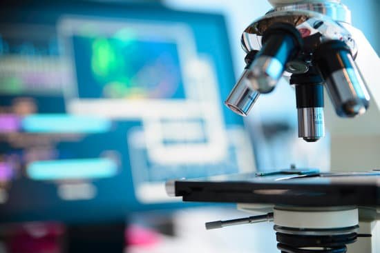
How has the microscope helped people? A microscope lets the user see the tiniest parts of our world: microbes, small structures within larger objects and even the molecules that are the building blocks of all matter. The ability to see otherwise invisible things enriches our lives on many levels.
How is microscope useful to us? A microscope is an instrument that is used to magnify small objects. Some microscopes can even be used to observe an object at the cellular level, allowing scientists to see the shape of a cell, its nucleus, mitochondria, and other organelles.
What have microscopes helped us discover? Microscopes allow humans to see cells that are too tiny to see with the naked eye. Therefore, once they were invented, a whole new microscopic world emerged for people to discover. On a microscopic level, new life forms were discovered and the germ theory of disease was born.
How did the invention of microscopes change the world? The invention of the microscope allowed scientists and scholars to study the microscopic creatures in the world around them. … The microscope allowed human beings to step out of the world controlled by things unseen and into a world where the agents that caused disease were visible, named and, over time, prevented.
How has the microscope helped people? – Related Questions
What is a picture taken through a microscope called?
A micrograph or photomicrograph is a photograph or digital image taken through a microscope or similar device to show a magnified image of an object. This is opposed to a macrograph or photomacrograph, an image which is also taken on a microscope but is only slightly magnified, usually less than 10 times.
What is the path of light in a compound microscope?
The path of light through a microscope. Modern microscopes are complex precision instruments. Light, originating in the light source (1), is focused by the condensor (2) onto the specimin (3). The light then enters the objective lens (4) and the image is magnified.
What does the ocular lens do in a microscope?
The image magnified by the objective lens is further magnified by the ocular lens for observation. An ocular lens consists of one to three lenses and is also provided with a mechanism, called a field stop, that removes unnecessary reflected light and aberration.
What do you use for microscopic photography?
Generally, an optical microscope is used in creating a micrograph or a photomicrograph. Most photographers, however, simply connect their digital camera to a microscope so that they can tale highly magnified photos of small-sized subjects.
Which microscope to use to see bacteria yeast?
Microscopes are essential for viewing cells and microorganisms. Anton van Leeuwenhoek was the first to observe human blood cells and to observe yeast and bacteria, which he referred to as animalcules. In this lab, you will use the compound light microscope to observe these same kinds of cells.
What does the objective lens do on a microscope simple?
The objective, located closest to the object, relays a real image of the object to the eyepiece. This part of the microscope is needed to produce the base magnification. The eyepiece, located closest to the eye or sensor, projects and magnifies this real image and yields a virtual image of the object.
What is the scanning objective lens on a microscope?
A scanning objective lens provides the lowest magnification power of all objective lenses. 4x is a common magnification for scanning objectives and, when combined with the magnification power of a 10x eyepiece lens, a 4x scanning objective lens gives a total magnification of 40x.
Why use alcohol to clean microscope?
Disinfecting after cleaning helps to lower the risk of spreading infection to other lab members. … Disinfecting the frame: We recommend applying 70% ethanol since it effectively disinfects the microscope without damaging the frame. Remember avoid using organic solvents except ethanol that may damage plastic parts.
Are microscopic thin walled vessels?
microscopic, thin-walled vessels that connect the smallest arteries to the smallest veins. Lines the inside of vessels and is exposed to the blood. the middle layer, usually the thickest as well. … Def: small vessels that link arterioles and capillaries.
What type of microscope should i use for hair?
A stereo microscope is typically used for the initial examination of hair (mounted and unmounted) before moving on to the compound microscope.
Can see the electrons by strongest microscope?
World’s Best Microscope Can Produce Images Less Than Diameter Of Single Hydrogen Atom. … TEAM 0.5 is the world’s most powerful transmission electron microscope and is capable of producing images with half-angstrom resolution, less than the diameter of a single hydrogen atom.
What type of microscope is used to view living cells?
Compound microscopes are light illuminated. The image seen with this type of microscope is two dimensional. This microscope is the most commonly used. You can view individual cells, even living ones.
What does microscopic hematuria mean?
“Microscopic” means something is so small that it can only be seen through a special tool called a microscope. “Hematuria” means blood in the urine. So, if you have microscopic hematuria, you have red blood cells in your urine. These blood cells are so small, though, you can’t see the blood when you urinate.
What was the source of microscopic animalcules described by leeuwenhoek?
In 1674 he likely observed protozoa for the first time and several years later bacteria. Those “very little animalcules” he was able to isolate from different sources, such as rainwater, pond and well water, and the human mouth and intestine.
What electron microscopes are used for?
Electron microscopes are used to investigate the ultra structure of a wide range of biological and inorganic specimens including microorganisms, cells, large molecules, biopsy samples, metals, and crystals. Industrially, electron microscopes are often used for quality control and failure analysis.
What cell organelles can be seen under a light microscope?
Note: The nucleus, cytoplasm, cell membrane, chloroplasts and cell wall are organelles which can be seen under a light microscope.
What microscope magnification to begin?
Why do you need to start with 4x in magnification on a microscope? The 4x objective lens has the lowest power and, therefore the highest field of view. As a result, it is easier to locate the specimen on the slide than if you start with a higher power objective.
What organelles can be seen under a light microscope?
Note: The nucleus, cytoplasm, cell membrane, chloroplasts and cell wall are organelles which can be seen under a light microscope.
What is the total magnification of a compound microscope?
Total magnification: In a compound microscope the total magnification is the product of the objective and ocular lenses (see figure below). The magnification of the ocular lenses on your scope is 10X. Immersion Oil: Clear, finely detailed images are achieved by contrasting the specimen with their medium.
When is an electron microscope used?
Electron microscopes are used to investigate the ultra structure of a wide range of biological and inorganic specimens including microorganisms, cells, large molecules, biopsy samples, metals, and crystals. Industrially, electron microscopes are often used for quality control and failure analysis.
Can we observe a typical virus with a light microscope?
Standard light microscopes allow us to see our cells clearly. However, these microscopes are limited by light itself as they cannot show anything smaller than half the wavelength of visible light – and viruses are much smaller than this.

