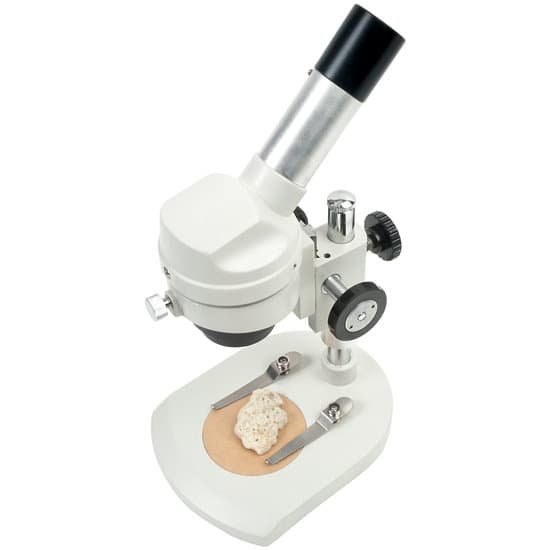
How have microscopes advanced science? A microscope allows scientists to view detailed relationships between the structures and functions at different levels of resolution. Microscopes have continued to be improved since they were first invented and used by early scientists like Anthony Leeuwenhoek to observe bacteria, yeast and blood cells.
Why are microscopes important in science? A microscope is an instrument that is used to magnify small objects. Some microscopes can even be used to observe an object at the cellular level, allowing scientists to see the shape of a cell, its nucleus, mitochondria, and other organelles.
What advances have been made to the microscope? In the 20th century, new instruments such as the electron microscope increased magnification and offered new insights into the body and disease, allowing scientists to see organisms such as viruses for the first time.
What magnification do you need to see mitochondria? b: The mitochondria (M) intermingled by rough endoplasmic reticulum (RER). The mitochondrial cristae are seen. Magnification: ×20,000.
How have microscopes advanced science? – Related Questions
Can you see colors with an electron microscope?
The reason is pretty basic: color is a property of light (i.e., photons), and since electron microscopes use an electron beam to image a specimen, there’s no color information recorded. The area where electrons pass through the specimen appears white, and the area where electrons don’t pass through appears black.
What does 40x mean on a microscope?
A 40x objective makes things appear 40 times larger than they actually are. Comparing objective magnification is relative—a 40x objective makes things twice as big as a 20x objective while a 60x objective makes them six times larger than a 10x objective. The eyepiece in a typical desktop microscope is 10x.
What kind of microscope can see organelles?
The electron microscope is necessary to see smaller organelles like ribosomes, macromolecular assemblies, and macromolecules. With light microscopy, one cannot visualize directly structures such as cell membranes, ribosomes, filaments, and small granules and vesicles.
What might a scientist use light microscope?
Light microscopes are extremely versatile instruments. They can be used to examine a wide variety of types of specimen, frequently with minimal preparation. Light microscopes can be adapted to examine specimens of any size, whole or sectioned, living or dead, wet or dry, hot or cold, and static or fast-moving.
What is retardation microscope?
The first order retardation plate is a standard accessory that is frequently utilized to determine the optical sign (positive or negative) of a birefringent specimen in polarized light microscopy. In addition, the retardation plate is also useful for enhancing contrast in weakly birefringent specimens.
What does the base do in a microscope?
Base: The bottom of the microscope, used for support Illuminator: A steady light source (110 volts) used in place of a mirror.
What are the four types of microscope?
There are several different types of microscopes used in light microscopy, and the four most popular types are Compound, Stereo, Digital and the Pocket or handheld microscopes. Some types are best suited for biological applications, where others are best for classroom or personal hobby use.
What does the arm do on a compound light microscope?
Arm connects to the base and supports the microscope head. It is also used to carry the microscope.
What are uses of microscopes?
A microscope is an instrument that is used to magnify small objects. Some microscopes can even be used to observe an object at the cellular level, allowing scientists to see the shape of a cell, its nucleus, mitochondria, and other organelles.
How have microscopes helped us to understand science?
Microscopes allow humans to see cells that are too tiny to see with the naked eye. Therefore, once they were invented, a whole new microscopic world emerged for people to discover. On a microscopic level, new life forms were discovered and the germ theory of disease was born.
How to clean used microscope slides?
When slides get soiled, you can clean them with soapy water or isopropyl alcohol. Do not immerse slides in water or soak them in it. This loosens the cover glass adhesive, causing the cover glass to come off and possibly ruin the slide.
What is a coma in a microscope?
Specifically, coma is defined as a variation in magnification over the entrance pupil. In refractive or diffractive optical systems, especially those imaging a wide spectral range, coma can be a function of wavelength, in which case it is a form of chromatic aberration.
What is the use of eyepiece in microscope?
The eyepiece, located closest to the eye or sensor, projects and magnifies this real image and yields a virtual image of the object. Eyepieces typically produce an additional 10X magnification, but this can vary from 1X – 30X. Figure 1 illustrates the components of a compound microscope.
What is electron microscopes used for?
Electron microscopy (EM) is a technique for obtaining high resolution images of biological and non-biological specimens. It is used in biomedical research to investigate the detailed structure of tissues, cells, organelles and macromolecular complexes.
What is the most commonly used microscope?
A compound microscope is the most common type of microscope used today, which mechanism is explained earlier. It is basically a microscope that has a lens or a camera on it that has a compound medium in between. This compound medium allows for magnifications in a very fine scale.
How to determine total magnification of a compound microscope?
To get the total magnification take the power of the objective (4X, 10X, 40x) and multiply by the power of the eyepiece, usually 10X.
Is a microscope upside down?
Microscopes invert images which makes the picture appear to be upside down. The reason this happens is that microscopes use two lenses to help magnify the image. Some microscopes have additional magnification settings which will turn the image right-side-up.
How does gram negative look under a microscope?
Gram negative bacteria appear a pale reddish color when observed under a light microscope following Gram staining. This is because the structure of their cell wall is unable to retain the crystal violet stain so are colored only by the safranin counterstain.
How to see live bacteria under microscope?
In order to see bacteria, you will need to view them under the magnification of a microscopes as bacteria are too small to be observed by the naked eye. Most bacteria are 0.2 um in diameter and 2-8 um in length with a number of shapes, ranging from spheres to rods and spirals.
Why was the microscope important in the renaissance?
The development of the microscope during the European Renaissance impacted both the ancient world and modern world by giving students something to learn in the classrooms and other scientists things to discover. Around year 1595 the microscope, one of the greatest inventions, was made.

