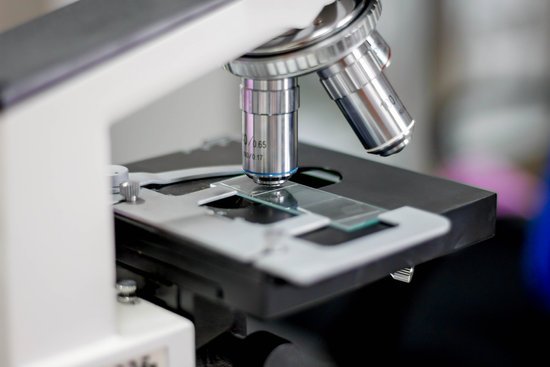
How is a scanning tunneling microscope etch? One of the most commonly used methods to create such a tip is electrochemical etching of a metal wire. … The wire is etched away at the meniscus of the solution, until the weight of the submerged wire is greater than the tensile force at the etching point. The wire then breaks at the meniscus and an STM tip is created.
What is a scanning tunneling microscope and how does it work? The scanning tunneling microscope (STM) works by scanning a very sharp metal wire tip over a surface. By bringing the tip very close to the surface, and by applying an electrical voltage to the tip or sample, we can image the surface at an extremely small scale – down to resolving individual atoms.
What are the components of scanning tunneling microscope? The main components of a scanning tunneling microscope are the scanning tip, piezoelectrically controlled height (z axis) and lateral (x and y axes) scanner, and coarse sample-to-tip approach mechanism. The microscope is controlled by dedicated electronics and a computer.
What is the purpose of a scanning tunneling microscope? The scanning tunneling microscope (STM) is widely used in both industrial and fundamental research to obtain atomic-scale images of metal surfaces.
How is a scanning tunneling microscope etch? – Related Questions
What type of microscope has highest resolution?
Out of all types of microscopes, the electron microscope has the greatest capability in achieving high magnification and resolution levels, enabling us to look at things right down to each individual atom.
How the microscope is used?
A microscope is an instrument that is used to magnify small objects. … It is through the microscope’s lenses that the image of an object can be magnified and observed in detail. A simple light microscope manipulates how light enters the eye using a convex lens, where both sides of the lens are curved outwards.
What kind of lenses do electron microscopes use?
Electron and ion microscopes use a beam of charged particles instead of light, and use electromagnetic or electrostatic lenses to focus the particles. They can see features as small as one-tenth of a nanometer (one ten billionth of a meter), including individual atoms.
Do we have a microscope that can see atoms?
An electron microscope can be used to magnify things over 500,000 times, enough to see lots of details inside cells. There are several types of electron microscope. A transmission electron microscope can be used to see nanoparticles and atoms.
What is the definition of arm on a microscope?
Arm. The part of the microscope that connects the eyepiece tube to the base. Articulated Arm Part of a boom microscope stand, an articulated arm has one or more joints to enable a greater variety of movement of the microscope head and, as a result, more versatile range of viewing options.
What are confocal microscopes used for?
The primary functions of a confocal microscope are to produce a point source of light and reject out-of-focus light, which provides the ability to image deep into tissues with high resolution, and optical sectioning for 3D reconstructions of imaged samples.
How does an sem microscope work?
The SEM is an instrument that produces a largely magnified image by using electrons instead of light to form an image. A beam of electrons is produced at the top of the microscope by an electron gun. … Once the beam hits the sample, electrons and X-rays are ejected from the sample.
What is the use of scanning electron microscope?
Scanning electron microscopy can help identify cracks, imperfections, or contaminants on the surfaces of coated products. Industries, like cosmetics, that work with tiny particles can also use scanning electron microscopy to learn more about the shape and size of the small particles they work with.
When were modern microscopes invented?
In the late 16th century several Dutch lens makers designed devices that magnified objects, but in 1609 Galileo Galilei perfected the first device known as a microscope.
What can scanning electron microscopes see cells?
Because of its great depth of focus, a scanning electron microscope is the EM analog of a stereo light microscope. It provides detailed images of the surfaces of cells and whole organisms that are not possible by TEM. It can also be used for particle counting and size determination, and for process control.
Where is the condenser diaphragm on a microscope?
On upright microscopes, the condenser is located beneath the stage and serves to gather wavefronts from the microscope light source and concentrate them into a cone of light that illuminates the specimen with uniform intensity over the entire viewfield.
What does the term resolution mean in relation to microscopes?
In microscopy, the term ‘resolution’ is used to describe the ability of a microscope to distinguish detail. In other words, this is the minimum distance at which two distinct points of a specimen can still be seen – either by the observer or the microscope camera – as separate entities.
What illuminates the specimen on a microscope?
Microscopes are designated as either light microscopes or electron microscopes. … The specimen is illuminated by a beam of tungsten light focused on it by a sub-stage lens called a condenser, and the result is that the specimen appears dark against a bright background.
How far can an electron microscope zoom?
This makes electron microscopes more powerful than light microscopes. A light microscope can magnify things up to 2000x, but an electron microscope can magnify between 1 and 50 million times depending on which type you use! To see the results, look at the image below.
What are the most powerful microscopes used?
Lawrence Berkeley National Labs just turned on a $27 million electron microscope. Its ability to make images to a resolution of half the width of a hydrogen atom makes it the most powerful microscope in the world.
What is the function of a dissecting microscope?
A dissecting microscope, also known as a stereo microscope, is used to perform dissection of a specimen or sample. It simply gives the person doing the dissection a magnified, 3-dimensional view of the specimen or sample so more fine details can be visualized.
What does sperm under a microscope look like?
The air-fixed, stained spermatozoa are observed under a bright-light microscope at 400x or 1000x magnification. Their viability and mor- phology can be analysed at the same time. Those appearing red-pink in colour have a damaged membrane whereas white sperm are viable, as in Photo 2.
What is pseudopods under a microscope?
Essentially, Pseudopodia are temporary projections of the cytoplasm that make it possible for amoebae to move. Pseudopods are some of the most distinguishable features of amoebae and their formation is based on the flow of the protoplasm.
What microscope is used to look at cells?
Two types of electron microscopy—transmission and scanning—are widely used to study cells. In principle, transmission electron microscopy is similar to the observation of stained cells with the bright-field light microscope.
When did the microscope come out?
The first compound microscopes date to 1590, but it was the Dutch Antony Van Leeuwenhoek in the mid-seventeenth century who first used them to make discoveries. When the microscope was first invented, it was a novelty item.
What colors are the parts of spirogyra under a microscope?
The cells in the filamentous structure are characterized by one or more spiral chloroplasts that give the characteristic green color to the organism.

