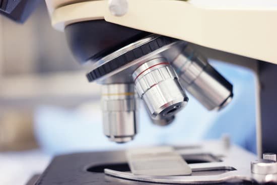
How is resolution of microscope measured? The resolution of an optical microscope is defined as the shortest distance between two points on a specimen that can still be distinguished by the observer or camera system as separate entities.
How do you determine the resolution of a microscope? Like the compound light, the resolution for a confocal microscope is about 1.2 nanometers. Scanning Electron Microscope Resolution: In a SEM, an electron beam scans rapidly over the surface of the sample specimen and yields an image of the topography of the surface. The resolution of a SEM is about 10 nanometers (nm).
What is the unit of resolution in microscope? The resolving power of an objective lens is measured by its ability to differentiate two lines or points in an object. The greater the resolving power, the smaller the minimum distance between two lines or points that can still be distinguished. The larger the N.A., the higher the resolving power.
What is the measure of the resolving power of a microscope? An optical system’s resolution can be measured by imaging the alternating light and dark lines at successively finer spatial scales, as displayed in Fig. 4. The spatial scale at which the line pairs become indistinguishable defines a resolution cut- off for a particular camera.
How is resolution of microscope measured? – Related Questions
What does electron microscopes do?
The electron microscope uses a beam of electrons and their wave-like characteristics to magnify an object’s image, unlike the optical microscope that uses visible light to magnify images.
What scientist made the first microscope?
The development of the microscope allowed scientists to make new insights into the body and disease. It’s not clear who invented the first microscope, but the Dutch spectacle maker Zacharias Janssen (b. 1585) is credited with making one of the earliest compound microscopes (ones that used two lenses) around 1600.
What are ocular lens on a microscope?
The ocular lenses are the lenses closest to the eye and usually have a 10x magnification. Since light microscopes use binocular lenses there is a lens for each eye.
Is it possible to see dna under microscope?
Given that DNA molecules are found inside the cells, they are too small to be seen with the naked eye. … While it is possible to see the nucleus (containing DNA) using a light microscope, DNA strands/threads can only be viewed using microscopes that allow for higher resolution.
Why use electron microscope?
Electrons have much a shorter wavelength than visible light, and this allows electron microscopes to produce higher-resolution images than standard light microscopes. Electron microscopes can be used to examine not just whole cells, but also the subcellular structures and compartments within them.
Can you focus eyepiece of a compound microscope?
The eyepiece, also called the ocular lens, is a low power lens. The objective lenses of compound microscopes are parfocal. You do not need to refocus (except for fine adjustment) when switching to a higher power if the object is in focus on a lower power. The field of view is widest on the lowest power objective.
What year was the compound microscope made?
The first compound microscopes date to 1590, but it was the Dutch Antony Van Leeuwenhoek in the mid-seventeenth century who first used them to make discoveries. When the microscope was first invented, it was a novelty item.
What part of the microscope makes small objects appear bigger?
A simple light microscope manipulates how light enters the eye using a convex lens, where both sides of the lens are curved outwards. When light reflects off of an object being viewed under the microscope and passes through the lens, it bends towards the eye. This makes the object look bigger than it actually is.
How can a microscope help a scientist use scientific methodology?
A new microscope is giving scientists a clearer, more comprehensive view of biological processes as they unfold in living animals. The microscope produces images of entire organisms, such as a zebrafish or fruit fly embryo, with enough resolution in all three dimensions that each cell appears as a distinct structure.
Will object be inverted in microscope?
Microscopes invert images which makes the picture appear to be upside down. The reason this happens is that microscopes use two lenses to help magnify the image.
What does diffuser do in microscope?
The light diffuser illuminates the specimen with substantially spatially isotropic light which passes through the stage evenly and without distinction as to direction producing an image for observation having substantially reduced diffraction shadows visible through the microscope which obscure the specimen.
How do you clean a microscope lens?
Dip a lens wipe or cotton swab into distilled water and shake off any excess liquid. Then, wipe the lens using the spiral motion. This should remove all water-soluble dirt.
What kind of light source for microscope with mirror?
A mirror illumination involves a small parabolic mirror that uses an outside light source (usually a light bulb on the ceiling or a desk lamp) to concentrate light up through the condenser lens and through the sample material.
What would be visible to a light microscope?
Explanation: You can see most bacteria and some organelles like mitochondria plus the human egg. You can not see the very smallest bacteria, viruses, macromolecules, ribosomes, proteins, and of course atoms.
What is the meaning dissecting microscope?
A dissecting microscope is used to view three-dimensional objects and larger specimens, with a maximum magnification of 100x. This type of microscope might be used to study external features on an object or to examine structures not easily mounted onto flat slides.
Is microscopic colitis contagious?
Colon inflammation caused by infection by a virus or bacteria can be spread, but autoimmune conditions causing colitis are not transmissible.
What do you use a light microscope for?
A light microscope is an optical instrument used to view objects too small to with the naked eye. It is so-called because it employs the use of white or visible light to illuminate the object of interest so it can be magnified and viewed through one or a series of lenses.
What is meant by scanning tunneling microscope?
: a microscope that makes use of the phenomenon of tunneling electrons to map the positions of individual atoms in a surface or to move atoms around on a surface. Other Words from scanning tunneling microscope Example Sentences Learn More About scanning tunneling microscope.
What can be seen by an electron microscope?
Some electron microscopes can detect objects that are approximately one-twentieth of a nanometre (10-9 m) in size – they can be used to visualise objects as small as viruses, molecules or even individual atoms.
Where is the diaphragm in a microscope?
Iris Diaphragm controls the amount of light reaching the specimen. It is located above the condenser and below the stage. Most high quality microscopes include an Abbe condenser with an iris diaphragm.
How have microscopes improved?
Microscopes became more stable and smaller. Lens improvements solved many of the optical problems that were common in earlier versions. The history of the microscope widens and expands from this point with people from around the world working on similar upgrades and lens technology at the same time.

