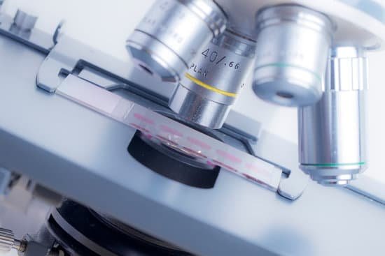
How light and electron microscopes work? Electron microscopes differ from light microscopes in that they produce an image of a specimen by using a beam of electrons rather than a beam of light. Electrons have much a shorter wavelength than visible light, and this allows electron microscopes to produce higher-resolution images than standard light microscopes.
How does an electron microscope work? The electron microscope uses a beam of electrons and their wave-like characteristics to magnify an object’s image, unlike the optical microscope that uses visible light to magnify images. … This stream is confined and focused using metal apertures and magnetic lenses into a thin, focused, monochromatic beam.
What are light and electron microscopes? There are two main types of microscope: light microscopes are used to study living cells and for regular use when relatively low magnification and resolution is enough. electron microscopes provide higher magnifications and higher resolution images but cannot be used to view living cells.
Who undertook the first definitive study of fingerprints as a method of identification? Fingerprinting replaced this procedure in the 1900’s. Bertillion also earned the distinction of being the father of criminal identification. Frances Galton undertook the first definitive study of fingerprints and developed a methodology of classifying them in 1892.
How light and electron microscopes work? – Related Questions
What can be seen under a dissection microscope?
With a dissecting microscope whole objects can be viewed in three dimensions. Samples do not need to be sliced, and larger, live animals can be observed. Light can be passed through from underneath the sample, but also from the top or side using an external light source.
Did robert hooke use a light microscope?
He designed his own light microscope, which used multiple glass lenses to light and magnify specimens. Under his microscope, Hooke examined a diverse collection of organisms. A gifted illustrator, he drew and explained what he saw. This record of his observations became Micrographia.
What types of specimens are viewed with a compound microscope?
Compound microscopes are used to view small samples that can not be identified with the naked eye. These samples are typically placed on a slide under the microscope. When using a stereo microscope, there is more room under the microscope for larger samples such as rocks or flowers and slides are not required.
What is the advantage of staining tissue before examining microscopically?
The most basic reason that cells are stained is to enhance visualization of the cell or certain cellular components under a microscope. Cells may also be stained to highlight metabolic processes or to differentiate between live and dead cells in a sample.
Which of these is defined as a microscopic living organism?
Technically a microorganism or microbe is an organism that is microscopic. The study of microorganisms is called microbiology. Microorganisms can be bacteria, fungi, archaea or protists.
How long has mankind been using microscopes?
In the late 16th century several Dutch lens makers designed devices that magnified objects, but in 1609 Galileo Galilei perfected the first device known as a microscope. Dutch spectacle makers Zaccharias Janssen and Hans Lipperhey are noted as the first men to develop the concept of the compound microscope.
Can i watch stomata with a microscope?
Most people use a traditional compound microscope at 400x to see individual stomata on plant leaves. It is possible to see stomata as small white dots on the underside of a leaf at magnifications from 50-100x, but you will not be able to do real stomatal counts, or see individual stomata.
What type of radiation does a light microscope use?
Radiation Type: Light microscopes use light (approx wavelength 400-700 nm), electron microscopes use beams of electrons (approx equivalent wavelength 1 nm). Control of image formation : Light via glass lenses, beams of electrons can be focused using electromagnets due to negative charge on electrons.
What is the meaning of base in microscope?
Base: The bottom of the microscope, used for support. Illuminator: A steady light source (110 volts) used in place of a mirror. If your microscope has a mirror, it is used to reflect light from an external light source up through the bottom of the stage.
Why do some microscopes invert images?
Microscopes invert images which makes the picture appear to be upside down. The reason this happens is that microscopes use two lenses to help magnify the image. Some microscopes have additional magnification settings which will turn the image right-side-up.
When can you see cells without a microscope?
The human eye cannot see most cells without the aid of a microscope. However, some large amoebas and bacteria, and some cells within complex multicellular organisms like humans and squid, can be viewed without aids.
Do you use light or electron microscope to see bacteria?
If you want to look at things like viruses, bacteria, or molecules passing through cell walls, you must use an electron microscope.
What are the uses of an electron microscope?
Electron microscopes are used to investigate the ultrastructure of a wide range of biological and inorganic specimens including microorganisms, cells, large molecules, biopsy samples, metals, and crystals. Industrially, electron microscopes are often used for quality control and failure analysis.
What is a good sentence for microscope?
1 Love looks with telescope; envy with microscope. 2 The microscope magnified the object two hundred times. 3 The microscope capacitates small objects to be observed. 4 An object was magnified 200 times by the microscope.
How do they age deer teeth with a microscope?
The slice of tooth is then placed on a slide and a special dye is added to enhance viewing. It is placed under a microscope. Lines within the tooth’s diameter are readily visible and can be counted much like the rings of growth on a tree, indicating a deer’s age.
What is meant by the compound microscope?
A compound microscope is a microscope that uses multiple lenses to enlarge the image of a sample. … Compound microscopes usually include exchangeable objective lenses with different magnifications (e.g 4x, 10x, 40x and 60x), mounted on a turret, to adjust the magnification.
What did scientist discover with the help of microscopes?
The invention of the microscope allowed scientists to see cells, bacteria, and many other structures that are too small to be seen with the unaided eye. It gave them a direct view into the unseen world of the extremely tiny.
What is the diaphragm used function in the microscope?
The field diaphragm controls how much light enters the substage condenser and, consequently, the rest of the microscope.
Which microscope is most commonly used in crime laboratories?
The stereoscopic microscope is the most frequently used and versatile microscope found in the crime laboratory. When you increase the compound microscope magnification its field of view decreases.
When and who invented the compound and electron microscope?
The invention of the electron microscope by Max Knoll and Ernst Ruska at the Berlin Technische Hochschule in 1931 finally overcame the barrier to higher resolution that had been imposed by the limitations of visible light. Since then resolution has defined the progress of the technology.

