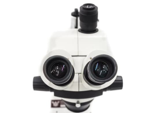
How the pinhole microscope works? A confocal microscope works with a laser and pinhole spatial filters. … The mirrors scan the laser across the sample. The dye and the emitted light get descanned by the mirrors that scan the excitation light. From there, the emitted light passes through the dichroic, then focuses on the pinhole.
What it is the function of the pinhole?
What is the purpose of pinhole aperture in confocal microscopy? The pinhole aperture also serves to eliminate much of the stray light passing through the optical system. Coupling of aperture-limited point scanning to a pinhole spatial filter at the conjugate image plane is an essential feature of the confocal microscope.
How does corneal confocal microscopy work? Corneal confocal microscopy is a novel clinical technique for the study of corneal cellular structure. It provides images which are comparable to in-vitro histochemical techniques delineating corneal epithelium, Bowman’s layer, stroma, Descemet’s membrane and the corneal endothelium.
How the pinhole microscope works? – Related Questions
How are refracting telescopes and simple light microscopes different?
Since telescopes view large objects — faraway objects, planets or other astronomical bodies — its objective lens produces a smaller version of the actual image. On the other hand, microscopes view very small objects, and its objective lens produces a larger version of the actual image.
Can a scanning electron microscope view living specimens?
Electron microscopes use a beam of electrons instead of beams or rays of light. Living cells cannot be observed using an electron microscope because samples are placed in a vacuum. … the scanning electron microscope (SEM) has a large depth of field so can be used to examine the surface structure of specimens.
What power microscope to see mushrooms?
To study fungal spores, basidia, cystidia, sphaerocysts and other tiny features of fungi you will need a microscope capable of at least x 400 magnification.
What does e coli look like under a microscope?
When viewed under the microscope, Gram-negative E. Coli will appear pink in color. The absence of this (of purple color) is indicative of Gram-positive bacteria and the absence of Gram-negative E. Coli.
What is microscopic anatomy quizlet?
microscopic anatomy. study of structures that cannot be seen without a microscope. cytology (microscopic anatomy) analyzes the structure of individual cells.
Who was the first person to make the microscope?
A Dutch father-son team named Hans and Zacharias Janssen invented the first so-called compound microscope in the late 16th century when they discovered that, if they put a lens at the top and bottom of a tube and looked through it, objects on the other end became magnified.
Is temperature a microscopic or macroscopic concept?
Sol: Temperature is a macroscopic concept . This means that temperature is an average property of the large number of molecules which constitute a system . We can not define the temperature of a single molecule .
What is the magnification of a stereo microscope?
The stereo- or dissecting microscope is an optical microscope variant designed for observation with low magnification (2 – 100x) using incident light illumination (light reflected off the surface of the sample is observed by the user), although it can also be combined with transmitted light in some instruments.
What should you do if you break a microscope slide?
If a slide or cover glass is broken, dispose of it and replace it immediately to prevent anyone from being cut. The adhesive used to attach a cover glass to a slide is applied as a liquid. As the liquid dries, it only hardens around the edges of the cover glass.
Can a nucleus be seen with a light microscope?
Thus, light microscopes allow one to visualize cells and their larger components such as nuclei, nucleoli, secretory granules, lysosomes, and large mitochondria. The electron microscope is necessary to see smaller organelles like ribosomes, macromolecular assemblies, and macromolecules.
What are magnification of all lens on microscope?
Most compound microscopes come with interchangeable lenses known as objective lenses. Objective lenses come in various magnification powers, with the most common being 4x, 10x, 40x, and 100x, also known as scanning, low power, high power, and (typically) oil immersion objectives, respectively.
How to view sperm under a microscope?
You can view sperm at 400x magnification. You do NOT want a microscope that advertises anything above 1000x, it is just empty magnification and is unnecessary. In order to examine semen with the microscope you will need depression slides, cover slips, and a biological microscope.
What kind of microscope do you need to see bacteria?
In order to actually see bacteria swimming, you’ll need a lens with at least a 400x magnification. A 1000x magnification can show bacteria in stunning detail.
How to determine magnifying power of microscope?
To figure the total magnification of an image that you are viewing through the microscope is really quite simple. To get the total magnification take the power of the objective (4X, 10X, 40x) and multiply by the power of the eyepiece, usually 10X.
What does n meningitidis look like under a microscope description?
Neisseria meningitidis is a Gram-negative, non-spore forming, non-motile, encapsulated, and non-acid-fast diplococci, which appears in kidney bean shape under the microscope.
What is the average magnification power of an electron microscope?
This makes electron microscopes more powerful than light microscopes. A light microscope can magnify things up to 2000x, but an electron microscope can magnify between 1 and 50 million times depending on which type you use!
What does a condenser in a microscope do?
On upright microscopes, the condenser is located beneath the stage and serves to gather wavefronts from the microscope light source and concentrate them into a cone of light that illuminates the specimen with uniform intensity over the entire viewfield.
What size microscope to see blood cells?
At 400x magnification you will be able to see bacteria, blood cells and protozoans swimming around. At 1000x magnification you will be able to see these same items, but you will be able to see them even closer up.
How do you find the magnification of a compound microscope?
In order to ascertain the total magnification when viewing an image with a compound light microscope, take the power of the objective lens which is at 4x, 10x or 40x and multiply it by the power of the eyepiece which is typically 10x.
When microscopes are parfocal magnification?
To simplify, if a compound microscope is parfocal, it means that when you change magnification sequentially (ex. 4x to 10x to 40x to 100x), it will only require a very slight turn of the fine focus knob with each increase or decrease to get the image in focus.
What kind of microscope can see bacteria?
On the other hand, compound microscopes are best for looking at all types of microbes down to bacteria. Some, however, are better than others. The magnification for most compound microscopes will be up to 1000X to 2500X.

