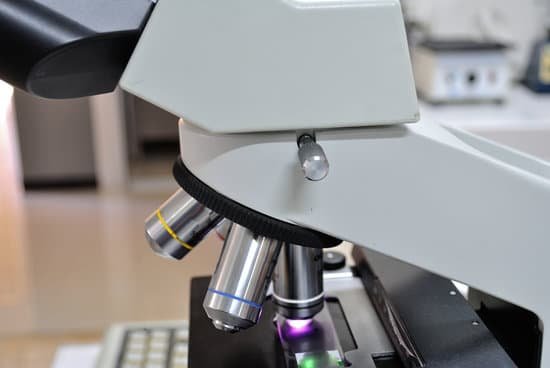
How to calibrate a microscope reticle? Calibrating a Microscope. To properly calibrate your reticle with a stage micrometer, align the zero line (beginning) of the stage micrometer with the zero line (beginning) of the reticle. Now, carefully scan over until you see the lines line up again.
How do you calibrate a microscope? To calibrate a reticle, switch to the scanning or low-power objective, place a clear plastic millimeter ruler on the stage, and then focus on the mm lines. What you see will resemble the illustrations below. Notice that microscope A is using an objective with a higher magnification than microscope B.
Who was the microscope invented by and when? Every major field of science has benefited from the use of some form of microscope, an invention that dates back to the late 16th century and a modest Dutch eyeglass maker named Zacharias Janssen.
Who invented the microscope in 1666? Antoni Van Leeuwenhoek (1635-1723) was a Dutch tradesman who became interested in microscopy while on a visit to London in 1666. Returning home, he began making simple microscopes of the sort that Robert Hooke had described in his, Micrographia, and using them to discover objects invisible to the naked eye.
How to calibrate a microscope reticle? – Related Questions
What microscope allows you to look at living specimens?
Compound microscopes are light illuminated. The image seen with this type of microscope is two dimensional. This microscope is the most commonly used. You can view individual cells, even living ones.
What is fine adjustment on a microscope?
FINE ADJUSTMENT KNOB — A slow but precise control used to fine focus the image when viewing at the higher magnifications.
Can dna be seen with microscopes?
Given that DNA molecules are found inside the cells, they are too small to be seen with the naked eye. For this reason, a microscope is needed. While it is possible to see the nucleus (containing DNA) using a light microscope, DNA strands/threads can only be viewed using microscopes that allow for higher resolution.
How to clean microscope objective?
Place the objective lens on a dust-free surface. 2. Gently blow away loose dust that is on the surface of the optical glass with a dust blower, as if any dust left on throughout the cleaning process could scratch the optical glass or coating. Blow the air across the lens surface to avoid damaging it.
How much does a new electron microscope cost?
The price of a new electron microscope can range from $80,000 to $10,000,000 depending on certain configurations, customizations, components, and resolution, but the average cost of an electron microscope is $294,000. The price of electron microscopes can also vary by type of electron microscope.
How much does a light microscope weigh?
Compound microscopes can range in weight from 3 pounds for a smaller compound microscope and up to 20 pounds for a lab grade microscope with a mounted camera.
How to calculate size of specimen under microscope?
Divide the number of cells in view with the diameter of the field of view to figure the estimated length of the cell. If the number of cells is 50 and the diameter you are observing is 5 millimeters in length, then one cell is 0.1 millimeter long. Measured in microns, the cell would be 1,000 microns in length.
What year was the first microscopes?
The first compound microscopes date to 1590, but it was the Dutch Antony Van Leeuwenhoek in the mid-seventeenth century who first used them to make discoveries. When the microscope was first invented, it was a novelty item.
Where is the eyepiece on a light microscope?
Eyepiece – also known as the ocular. this is the part used to look through the microscope. Its found at the top of the microscope. Its standard magnification is 10x with an optional eyepiece having magnifications from 5X – 30X.
What is microscopic hematuria causes?
The most common causes of microscopic hematuria are urinary tract infection, benign prostatic hyperplasia, and urinary calculi. However, up to 5% of patients with asymptomatic microscopic hematuria are found to have a urinary tract malignancy.
How to manipulate compound microscope?
Look at the objective lens (3) and the stage from the side and turn the focus knob (4) so the stage moves upward. Move it up as far as it will go without letting the objective touch the coverslip. Look through the eyepiece (1) and move the focus knob until the image comes into focus.
What is a binocular microscope mean?
n. A microscope having two eyepieces, one for each eye, so that the object can be viewed with both eyes.
Who is credited with making the first microscope?
The development of the microscope allowed scientists to make new insights into the body and disease. It’s not clear who invented the first microscope, but the Dutch spectacle maker Zacharias Janssen (b. 1585) is credited with making one of the earliest compound microscopes (ones that used two lenses) around 1600.
What do you use a microscope for in science?
A microscope is an instrument that is used to magnify small objects. Some microscopes can even be used to observe an object at the cellular level, allowing scientists to see the shape of a cell, its nucleus, mitochondria, and other organelles.
Is looking in microscope for long time bad?
Microscopes can cause eye strain from squinting and staring for too long. They can also cause back pain from hunching over and looking into the microscope. The ambient light and the magnification used in microscopes can also cause eye strain over time and can lead to long-term pain or damage to your eyes.
Can i see sperm under a microscope?
A semen microscope or sperm microscope is used to identify and count sperm. … You can view sperm at 400x magnification. You do NOT want a microscope that advertises anything above 1000x, it is just empty magnification and is unnecessary.
What is the resolving power of a standard light microscope?
For a light microscope, the highest practicable NA is around 1.4. For white light (lambda is approximately 0.53 m, the resolving power is 0.231 m, or 231nm.
Can a electron microscope microscope see bacteria?
Scanning electron microscopy allows you to see the outside structure of bacteria in detail (Fig. 3.9) and can be used, for example, to see whether an antimicrobial agent has an impact on cell structure or integrity.
How to calculate magnification using a light microscope?
To calculate the total magnification of the compound light microscope multiply the magnification power of the ocular lens by the power of the objective lens. For instance, a 10x ocular and a 40x objective would have a 400x total magnification. The highest total magnification for a compound light microscope is 1000x.
How to make a sample preparation for microscopic examination?
Mechanical preparation is a commonly used technique for preparing metallographic samples for microscopic analysis. It involves using abrasive particles in successively finer steps to strip material from the surface until achieving the desired result.

