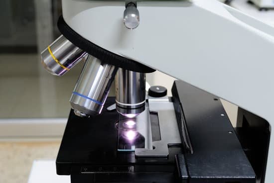
How to carry a compound light microscope? When carrying the light microscope, handlers must put one hand on the base at all times, to avoid dropping it, while the other hand should be on the arm. The microscope must never be carried upside down, since the ocular will fall out. It should never be swung when it is carried, according to Miami University.
How should you carry a compound light microscope? Always keep your microscope covered when not in use. Always carry a microscope with both hands. Grasp the arm with one hand and place the other hand under the base for support.
How do you carry a microscope from one place to another? The proper way to carry a microscope is to grasp the arm of the microscope with your dominant hand, lift the microscope up slowly, and use your other hand to firmly hold the base to stabilize the microscope as you transport it from one place to another.
Which two parts should you hold when carrying a compound light microscope? To carry the microscope, grasp the microscope’s arm with one hand. Place your other hand under the base. 2. Place the microscope on a table with the arm toward you.
How to carry a compound light microscope? – Related Questions
What is a microscope slide cover?
A microscope slide is a thin sheet of glass used to hold objects for examination under a microscope. … This smaller sheet of glass, called a cover slip or cover glass, is usually between 18 and 25 mm on a side.
What is resolution on microscope definition?
In microscopy, the term ‘resolution’ is used to describe the ability of a microscope to distinguish detail. In other words, this is the minimum distance at which two distinct points of a specimen can still be seen – either by the observer or the microscope camera – as separate entities.
What is high power on a compound microscope?
High power microscopes – also called compound microscopes because of their two-stage magnification – are the ‘typical’ microscope that is seen in laboratories and on television. High power microscopes typically magnify in the range ×40 to ×1000; much greater than this will only produce fuzzy images with no more detail.
Does microscope light damage the eyes?
For instance, long exposures to the operating microscope might result into photochemical retinal damage to the patient, a phenomenon recognized since the eighties of the previous century. The endoscope is potentially dangerous too, in particular due to the hardly accurately adjustable distance to the retina.
What does a transmission electron microscope cost?
The cost of a transmission electron microscope (TEM) can range from $300,000 to $10,000,000. The cost of a focused ion beam electron microscope (FIB) can range from $500,000 to $4,000,000. There can be a high degree of variation in the cost of an electron microscope between manufacturers and models.
Who discovered microscope in which year?
Lens Crafters Circa 1590: Invention of the Microscope. Every major field of science has benefited from the use of some form of microscope, an invention that dates back to the late 16th century and a modest Dutch eyeglass maker named Zacharias Janssen.
Can you see an atom with a light microscope?
The wavelength of visible light is about ten thousand times the length of a typical atom. … Since an atom is so much smaller than the wavelength of visible light, it’s much too small to change the way light is reflected, so observing an atom with an optical microscope will not work.
What does the condenser diaphragm control on a microscope?
Opening and closing of the condenser aperture diaphragm controls the angle of the light cone reaching the specimen. The setting of the condenser’s aperture diaphragm, along with the aperture of the objective, determines the realized numerical aperture of the microscope system.
How to identify malaria under microscope?
Malaria parasites can be identified by examining under the microscope a drop of the patient’s blood, spread out as a “blood smear” on a microscope slide. Prior to examination, the specimen is stained (most often with the Giemsa stain) to give the parasites a distinctive appearance.
What is the cost of a compound light microscope?
The most popular compound microscopes from some of the most well-known brands cost on average around $900-$1,200, although there are beginner microscopes that are just above the toy level that cost $100.
What does a rheostat do on a microscope?
rheostat – alters the current applied to the lamp to control the intensity of the light produced condenser – lens system that aligns and focuses the light from the lamp onto the specimen diaphragms or pinhole apertures – placed in the light path to alter the amount of light that reaches the condenser (for enhancing …
How much is an electron microscope?
The price of a new electron microscope can range from $80,000 to $10,000,000 depending on certain configurations, customizations, components, and resolution, but the average cost of an electron microscope is $294,000. The price of electron microscopes can also vary by type of electron microscope.
What is the arm of a microscope function?
Arm connects to the base and supports the microscope head. It is also used to carry the microscope.
When would i use a scanning electron microscope?
Geological sampling using a scanning electron microscope can determine weathering processes and morphology of the samples. Backscattered electron imaging can be used to identify compositional differences, while composition of elements can be provided by microanalysis.
What is the resolution power of electron microscope?
The resolution limit of electron microscopes is about 0.2nm, the maximum useful magnification an electron microscope can provide is about 1,000,000x.
What type of microscope is used to view insects?
Typically a low power stereo microscope is best for viewing insects because it will provide a 3D image. Ant as seen under a dissecting microscope. Bald faced hornet under the stereo microscope. Once you view the insects under a dissecting microscope, you may wish to view more details of a specific part of the insect.
Who was the first to see cells under a microscope?
Initially discovered by Robert Hooke in 1665, the cell has a rich and interesting history that has ultimately given way to many of today’s scientific advancements.
How does a dissection microscope work?
The stereo, stereoscopic or dissecting microscope is an optical microscope variant designed for low magnification observation of a sample, typically using light reflected from the surface of an object rather than transmitted through it.
Who invented the microscope in the scientific revolution?
Zacharias Janssen was a Dutch spectacle-maker from Middelburg associated Who invented the first optical telescope. Janssen is sometimes also credited for inventing the first truly compound microscope. Zacharias Janssen is generally believed to be the first investigator to invent the compound microscope.
What can be done with a microscope?
A microscope is an instrument that can be used to observe small objects, even cells. The image of an object is magnified through at least one lens in the microscope. This lens bends light toward the eye and makes an object appear larger than it actually is.
What does hyphae look like under microscope?
Hyphae are described as “gloeoplerous” (“gloeohyphae”) if their high refractive index gives them an oily or granular appearance under the microscope. These cells may be yellowish or clear (hyaline). They can sometimes selectively be coloured by sulphovanillin or other reagents.

