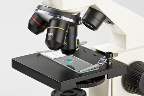
How to clean microscope cover slips? Wash the coverslips extensively in distilled water (two times), then double distilled water (two times). Be sure to wash out the acid between sticked coverslips. Rinse coverslips in 100% ethanol and dry them between sheets of whatman filter paper. Keep coverslip in a clean container.
How do you clean a microscope slide cover? Place all dirty microscope slides in a water basin full of warm water and detergent. … This will leave a detergent residue on the slides that will be difficult to remove. Wrap cleaned slides in sheets of clean paper until they are ready to be used again.
Can you wash microscope slides? After cleaning your coverslips and giving them a preliminary sterilization in 70% ethanol, pop them under the germicidal UV light of your tissue culture hood for 15 minutes and voilà! you have very sterile coverslips. Incubations under the UV light vary from 15 minutes, up to 1 or 4 hours, or even overnight.
How do you disinfect cover slips? If you’re using cover slips, you should also wash those between every use. Johnny L. The common type of microscope slides are the simple glass ones used for compound light microscopes, and yes, they can be used repeatedly. Just make sure you wash and dry the slide very well between each use.
How to clean microscope cover slips? – Related Questions
Who discovered stereo microscope?
In the early 1890’s, an American biologist and instrument maker, Horatio S. Greenough developed a stereo microscope which was an alternative design to the CMO microscope.
Can you see cells with a compound microscope?
Compound microscopes are light illuminated. The image seen with this type of microscope is two dimensional. This microscope is the most commonly used. You can view individual cells, even living ones.
How did the invention of microscopes help scientists?
The invention of the microscope allowed scientists to see cells, bacteria, and many other structures that are too small to be seen with the unaided eye. It gave them a direct view into the unseen world of the extremely tiny.
What happens to a scratched microscope objective?
They will scratch the delicate anti-reflection coatings on the front lens of the objective, and will ruin it, permanently causing flare and reduced contrast and reduced signal-to-noise ratio in the image.
How is a scanning electron microscope image formed?
The scanning electron microscope (SEM) produces images by scanning the sample with a high-energy beam of electrons. As the electrons interact with the sample, they produce secondary electrons, backscattered electrons, and characteristic X-rays.
What to use to clean lenses on compound light microscope?
To clean the eyepiece lens of your microscope, breathe onto the eyepiece lens and then wipe with lens tissue. For dirt that is difficult to remove, add ethanol (methanol in extreme cases) to a cotton swab, wipe the surface and then dry with a dry swab.
How are telescopes and microscopes similar?
Microscopes and telescopes are quite similar in that they are both utilized to view objects up close. … While microscopes provide the user with a view of material in an easier manner than the telescope user, since telescope use takes patience to find various objects in the sky.
How much are electron microscope?
The price of a new electron microscope can range from $80,000 to $10,000,000 depending on certain configurations, customizations, components, and resolution, but the average cost of an electron microscope is $294,000. The price of electron microscopes can also vary by type of electron microscope.
What is an electron microscope gcse?
Electron microscopes use a beam of electrons instead of beams or rays of light. … the transmission electron microscope (TEM) is used to examine thin slices or sections of cells or tissues. the scanning electron microscope (SEM) has a large depth of field so can be used to examine the surface structure of specimens.
What type of microscope to see bacteria in water?
In order to actually see bacteria swimming, you’ll need a lens with at least a 400x magnification. A 1000x magnification can show bacteria in stunning detail.
How to clean microscope stage?
Using a lint-free cotton swab, dip the end into your cleaning solution; alcohol,etc. Shake off excess fluid from the swab. Using the cotton end of the stick, start at the center of the lens using a circular motion and work your way to the outer edge. Gently wipe off any excess liquid with another dry lint-free swab.
How to see bacteria under light microscope?
Bacteria are difficult to see with a bright-field compound microscope for several reasons: They are small: In order to see their shape, it is necessary to use a magnification of about 400x to 1000x. The optics must be good in order to resolve them properly at this magnification.
Does electron microscope take pictures in color?
Why do electron microscopes produce black and white images? The reason is pretty basic: color is a property of light (i.e., photons), and since electron microscopes use an electron beam to image a specimen, there’s no color information recorded.
How to focus a microscope using the high power lens?
When focusing on a slide, ALWAYS start with either the 4X or 10X objective. Once you have the object in focus, then switch to the next higher power objective. Re-focus on the image and then switch to the next highest power.
How to do microscope magnification?
To figure the total magnification of an image that you are viewing through the microscope is really quite simple. To get the total magnification take the power of the objective (4X, 10X, 40x) and multiply by the power of the eyepiece, usually 10X.
What is the numerical aperture of a light microscope?
The numerical aperture of a microscope objective is the measure of its ability to gather light and to resolve fine specimen detail while working at a fixed object (or specimen) distance.
Can paper microscopes see blood cells?
The flat paper microscopes, about the size of an standard envelope, are inexpensive, portable, easy to assemble, and have a magnification of 140X and a 2-micron resolution, meaning users can see things as small as bacteria, blood cells and single-celled organisms.
Why do we call it a compound light microscope?
A compound microscope is a microscope that uses multiple lenses to enlarge the image of a sample. … The total magnification is calculated by multiplying the magnification of the ocular lens by the magnification of the objective lens.
Who developed the first hand lens and microscope?
It was Antony Van Leeuwenhoek (1632-1723), a Dutch draper and scientist, and one of the pioneers of microscopy who in the late 17th century became the first man to make and use a real microscope. He made his own simple microscopes, which had a single lens and were hand-held.
How many power can a light microscope have?
Magnification. The actual power or magnification of a compound optical microscope is the product of the powers of the ocular (eyepiece) and the objective lens. The maximum normal magnifications of the ocular and objective are 10× and 100× respectively, giving a final magnification of 1,000×.
What are light microscopes used to observe?
light microscopes are used to study living cells and for regular use when relatively low magnification and resolution is enough. electron microscopes provide higher magnifications and higher resolution images but cannot be used to view living cells.

