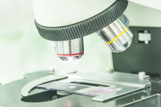
How to clean prepared microscope slides? When slides get soiled, you can clean them with soapy water or isopropyl alcohol. Do not immerse slides in water or soak them in it. This loosens the cover glass adhesive, causing the cover glass to come off and possibly ruin the slide.
How long do prepared microscope slides last? Used and stored in slide storage boxes properly, these prepared slides will last 5+ years with regular use.
How do you clean sliding slips covers? Wash the coverslips extensively in distilled water (two times), then double distilled water (two times). Be sure to wash out the acid between sticked coverslips. Rinse coverslips in 100% ethanol and dry them between sheets of whatman filter paper. Keep coverslip in a clean container.
How do you sterilize glass slides? Most slides and glass surfaces can be effectively cleaned with soapy water. Prepare a bath of soapy water. Use a lint-free wipe and cotton swab to gently rub the surface clean of dirt and residues. Rinse thoroughly in DI water and blow dry with nitrogen.
How to clean prepared microscope slides? – Related Questions
What color are endospores under a microscope?
Examine the slide under microscope for the presence of endospores. Endospores are bright green and vegetative cells are brownish red to pink.
What is the field of view in a compound microscope?
Field of view microscope definition in simple terms it is the area you see under the microscope for a particular magnification. Say, for example, you are viewing a cell or specimen under an optical microscope. The diameter of the circle that you see is the field of view of the microscope.
What power microscope to see blood cells?
At 400x magnification you will be able to see bacteria, blood cells and protozoans swimming around. At 1000x magnification you will be able to see these same items, but you will be able to see them even closer up.
What are some microscopic organisms?
Algae — these are single celled plants also known as phytoplankton (from the Greek, meaning drifting plants). Protozoa — these are single celled animals also known as zooplankton (from the Greek, meaning drifting animals). Bacteria — the most abundant organisms on earth.
How has the microscope changed our lives?
A microscope lets the user see the tiniest parts of our world: microbes, small structures within larger objects and even the molecules that are the building blocks of all matter. The ability to see otherwise invisible things enriches our lives on many levels.
How to handle a microscope properly?
Hold the microscope with one hand around the arm of the device, and the other hand under the base. This is the most secure way to hold and walk with the microscope. Avoid touching the lenses of the microscope. The oil and dirt on your fingers can scratch the glass.
Why do you carry a microscope by the base?
However, microscopes are extremely expensive, so you want to make sure you handle the device properly. Hold the microscope with one hand around the arm of the device, and the other hand under the base. This is the most secure way to hold and walk with the microscope. Avoid touching the lenses of the microscope.
What is the history of the optical microscope?
In the late 13th century, in Italy, clear silicate glass was produced for the very first time. As a result, both concave and convex lenses of relatively high quality started to be manufactured, paving the way for the construction of the first simple microscopes.
What are the three types of electron microscopes?
There are several different types of electron microscopes, including the transmission electron microscope (TEM), scanning electron microscope (SEM), and reflection electron microscope (REM.)
How does a microscope magnify an object?
A microscope is an instrument that can be used to observe small objects, even cells. The image of an object is magnified through at least one lens in the microscope. This lens bends light toward the eye and makes an object appear larger than it actually is.
What’s the iris diaphragm on a microscope?
Iris Diaphragm controls the amount of light reaching the specimen. It is located above the condenser and below the stage. Most high quality microscopes include an Abbe condenser with an iris diaphragm. Combined, they control both the focus and quantity of light applied to the specimen.
What is a microscopic colitis?
Microscopic colitis is a chronic inflammatory bowel disease (IBD) in which abnormal reactions of the immune system cause inflammation of the inner lining of your colon. Anyone can develop microscopic colitis, but the disease is more common in older adults and in women.
How to estimate field of view on a microscope?
For instance, if your eyepiece reads 10X/22, and the magnification of your objective lens is 40. First, multiply 10 and 40 to get 400. Then divide 22 by 400 to get a FOV diameter of 0.055 millimeters.
How to focus on high power microscope?
When focusing on a slide, ALWAYS start with either the 4X or 10X objective. Once you have the object in focus, then switch to the next higher power objective. Re-focus on the image and then switch to the next highest power.
How does an ultrasonic microscope work?
In contrast to this, the scanning acoustic microscope is a sequential imaging system in which a piezoelectric transducer emits a focussed ultrasound beam that propagates through a water, to the sample. The beam is scattered by the sample, and the scattered ultrasound wave is detected piezoelectrically.
Which microscope is 3 dimensional?
Scanning electron microscopy (SEM) is a powerful technique, traditionally used for imaging the surface of cells, tissues and whole multicellular organisms (see An Introduction to Electron Microscopy for Biologists)(Fig. 1).
What is the meaning of coarse adjustment on a microscope?
COARSE ADJUSTMENT KNOB — A rapid control which allows for quick focusing by moving the objective lens or stage up and down. It is used for initial focusing.
Why are images observed under light microscopes reversed and inverted?
The letter appears upside down and backwards because of two sets of mirrors in the microscope. This means that the slide must be moved in the opposite direction that you want the image to move. … These slides are thick, so they should only be viewed under low power.
Why do scientists use light microscopes?
One of two main types of microscopes and they use light and lenses to enlarge an image of an object. A simple light microscope has only one lens. Light Microscopes can enlarge images up to 1,500 times their original size. … Forensic scientists use microscopes to study evidence from crime scenes.
How do the stomata look under the microscope?
When viewed under the microscope, it’s possible to see the epidermal cells that tend to be irregular. In addition to the epidermal cells, one will also see the leaf spores (stomata) in between the epidermal cells. Typically, the stomata are bean shaped and will appear denser (darker) under the microscope.
What does the high power objective of a microscope do?
The high-powered objective lens (also called “high dry” lens) is ideal for observing fine details within a specimen sample. The total magnification of a high-power objective lens combined with a 10x eyepiece is equal to 400x magnification, giving you a very detailed picture of the specimen in your slide.

