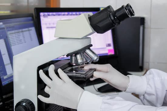
How to collect dust mites for microscope? Put the tape on the slide under the lens of the microscope with the power set to at least 10x magnification. Dust mites are 0.3 millimetres (0.012 in) in size, so they can’t be seen with the naked eye.
How do you find dust mites on a microscope? As I mentioned earlier, dust mites are microscopic creatures which cannot be seen by a naked human eye. However, they can easily be seen under a microscope with at least a 10x magnification lens. Most standard microscopes have 10x magnification eyepieces.
How do you get a dust mite sample? Dust samples for dust mite allergens can be collected using a dust cassette or by using a specially designed dust trap attached to a vacuum cleaner. A filter or a 9 square inch bed linen can otherwise be placed between the hose and the attachment of vacuum cleaner to collect the dust.
Can I see dust mites with a magnifying glass? No, you can’t see dust mites. They are so small (0.3mm) that you need a magnifying glass to see them. … The main food source for dust mites is dead skin from humans and animals. For this reason, if you don’t have a good solution for dust mites, they will be in your home permanently.
How to collect dust mites for microscope? – Related Questions
How to measure total magnification of a microscope?
To calculate the total magnification of the compound light microscope multiply the magnification power of the ocular lens by the power of the objective lens. For instance, a 10x ocular and a 40x objective would have a 400x total magnification. The highest total magnification for a compound light microscope is 1000x.
How do i connect my usb microscope to the television?
Plug the device into any open USB port on the computer or the television. Hold the microscope and lightly touch the lens to the specimen. The image should now be visible on the monitor or television screen.
What to do with a microscope?
A microscope is an instrument that can be used to observe small objects, even cells. The image of an object is magnified through at least one lens in the microscope. This lens bends light toward the eye and makes an object appear larger than it actually is.
What is the highest a light microscope can magnify?
Throughout their development, the magnification of light microscopes has increased, but very high magnifications are not possible. The maximum magnification with a light microscope is around ×1500.
What are stage controls on a microscope?
These allow you to move your slide while you are viewing it, but only if the slide is properly clipped in with the stage clips. Always find where these are on your microscope before you start viewing your slide.
Can you test for tb under a microscope?
In many countries, sputum smear microscopy remains the primary tool for the laboratory diagnosis of tuberculosis. It requires simple laboratory facilities, and when performed correctly, has a role in rapidly identifying infectious cases.
What are the 3 types of electron microscopes?
There are several different types of electron microscopes, including the transmission electron microscope (TEM), scanning electron microscope (SEM), and reflection electron microscope (REM.)
How to use a microscope and oil immersion?
Place “one drop” of immersion oil directly onto your coverslip. Slowly rotate your oil objective lens into place and bring the nose of your objective in contact with the drop of oil. Use only the fine focus control, very slowly bring your specimen back into focus.
What is microscopic colitis?
Microscopic colitis is a chronic inflammatory bowel disease (IBD) in which abnormal reactions of the immune system cause inflammation of the inner lining of your colon. Anyone can develop microscopic colitis, but the disease is more common in older adults and in women.
Which of these microscopes has the highest resolution?
Out of all types of microscopes, the electron microscope has the greatest capability in achieving high magnification and resolution levels, enabling us to look at things right down to each individual atom.
What microscope do biologists use?
Microscopes are especially useful in biology, where many biologist study organisms too small to see without help. They may use stereoscopes, compound microscopes, confocal microscopes, electron microscopes, or any of the specialized microscopes within each category.
What do microscope light parts do?
The optical parts of the microscope are used to view, magnify, and produce an image from a specimen placed on a slide.
Can microscopes be thrown away?
Examples of e-waste that should NOT be disposed of in your trash bin include: Kitchen equipment: Toasters, coffee makers, microwave ovens. Laboratory equipment: Hot plates, microscopes, calorimeters.
How long can microscopic colitis last?
The outlook for people with Microscopic Colitis is generally good. Four out of five can expect to be fully recovered within three years, with some even recovering without treatment. However, for those who experience persistent or recurrent diarrhea, long term budesonide may be necessary.
What is the iris diaphragm of a microscope?
: an adjustable diaphragm of thin opaque plates that can be turned by a ring so as to change the diameter of a central opening usually to regulate the aperture of a lens (as in a microscope)
How many times can a scanning electron microscope magnify?
SEM: magnifies 5 to ~ 500,000 times; sharp images of surface features. STEM: magnifies 5 to ~50 million times; the specimen appears flat.
How you calculate field size on a microscope?
For instance, if your eyepiece reads 10X/22, and the magnification of your objective lens is 40. First, multiply 10 and 40 to get 400. Then divide 22 by 400 to get a FOV diameter of 0.055 millimeters.
Who discovered microscope image?
In the late 16th century several Dutch lens makers designed devices that magnified objects, but in 1609 Galileo Galilei perfected the first device known as a microscope. Dutch spectacle makers Zaccharias Janssen and Hans Lipperhey are noted as the first men to develop the concept of the compound microscope.
How did zacharias janssen microscope work?
The early Janssen microscopes were compound microscopes, which use at least two lenses. The objective lens is positioned close to the object and produces an image that is picked up and magnified further by the second lens, called the eyepiece.
Is the physical chemical and microscopic examination of urine?
Urinalysis is the physical, chemical, and microscopic examination of urine. It involves a number of tests to detect and measure various compounds that pass through the urine.
Is the microscopic study of cells?
“cell”) are professionals who study cells via microscopic examinations and other laboratory tests. They are trained to determine which cellular changes are within normal limits and which are abnormal.

