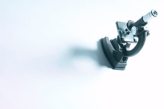
How to control the diaphragm on a microscope? Below. The light source is housed in the base of the microscope. It passes through the field iris diaphragm. The size of the field diaphragm is controlled by rotating a knurled ring which is concentric with it.
How do I control my iris diaphragm? You can adjust the diaphragm by turning it clockwise to close it, or counterclockwise to open it. Only open the iris diaphragm of the microscope to a point where the light passing through barely extends beyond the microscope’s field of view.
Why do you need to adjust the diaphragm on a microscope? SINCE SOMEONE ELSE IN ANOTHER LAB SECTION WILL BE USING YOUR MICROSCOPE IT MAY BE NECESSARY TO PERFORM THIS ADJUSTMENT EACH TIME YOU USE THE MICROSCOPE. DOING THIS WILL HELP PREVENT EYESTRAIN. ONCE THE SPECIMEN IS IN FOCUS, IT IS TIME TO ADJUST THE CONDENSER DIAPHRAGM APERTURE.
How should the condenser and diaphragm be adjusted for viewing? How should the condenser and diaphragm be adjusted for optimum viewing? Condenser is kept at highest point, just below stage. Diaphragm varies based on how much light is needed. Explain how to properly clean the lenses on a microscope.
How to control the diaphragm on a microscope? – Related Questions
Can molecules be seen under a microscope?
We can appreciate cells and bacteria with optical microscopes, while viruses and molecules are visible only under an electron microscope.
What is the magnification of the eyepiece on a microscope?
The standard eyepiece magnifies 10x. Check the objective lens of the microscope to determine the magnification, which is usually printed on the casing of the objective.
What happens if microscopic glass particles get into your lungs?
When workers inhale crystalline silica (dust), the lung tissue reacts by developing fibrotic nodules and scarring around the trapped silica particles. This fibrotic condition of the lung is called silicosis. If the nodules grow too large, breathing becomes difficult and death may result.
What is an objective in a microscope?
Introduction. The most important imaging component in the optical microscope is the objective, a complex multi-lens assembly that focuses light waves originating from the specimen and forms an intermediate image that is subsequently magnified by the eyepieces.
What does the microscope base do?
Base: The bottom of the microscope, used for support Illuminator: A steady light source (110 volts) used in place of a mirror.
How to look pollen under microscope?
When viewed under the stereo microscope, pollen grains will appear as grossly shaped, irregular structures/particles. However, the shape and appearance of the grains will vary depending on the type of pollen under investigation. For untreated grains, there is poor contrast compared to treated pollen grains.
How do microscope objectives magnify?
The object is brought to twice the focal distance in front of the lens. … Such finite tube length objectives project a real, inverted, and magnified image into the body tube of the microscope. This image comes into focus at the plane of the fixed diaphragm in the eyepiece.
What is the limited resolution of a microscope?
The resolution of the light microscope cannot be small than the half of the wavelength of the visible light, which is 0.4-0.7 µm. When we can see green light (0.5 µm), the objects which are, at most, about 0.2 µm.
What is the index of refraction of the plastic microscope?
Most plastics have refractive indices in the range from 1.3 to 1.7, but some high-refractive-index polymers can have values as high as 1.76.
How to figure out magnification on a microscope?
To figure the total magnification of an image that you are viewing through the microscope is really quite simple. To get the total magnification take the power of the objective (4X, 10X, 40x) and multiply by the power of the eyepiece, usually 10X.
How to determine the magnification produced by a microscope?
Total Magnification: To figure the total magnification of an image that you are viewing through the microscope is really quite simple. To get the total magnification take the power of the objective (4X, 10X, 40x) and multiply by the power of the eyepiece, usually 10X.
What do microscopes do to help us?
A microscope lets the user see the tiniest parts of our world: microbes, small structures within larger objects and even the molecules that are the building blocks of all matter. The ability to see otherwise invisible things enriches our lives on many levels.
What are the limitations of electron microscopes?
The main disadvantages are cost, size, maintenance, researcher training and image artifacts resulting from specimen preparation. This type of microscope is a large, cumbersome, expensive piece of equipment, extremely sensitive to vibration and external magnetic fields.
Can light microscopes see bacteria?
Generally speaking, it is theoretically and practically possible to see living and unstained bacteria with compound light microscopes, including those microscopes which are used for educational purposes in schools.
What is used to explain microscopic phenomena?
Classical physics is inadequate to handle microscopic domain and Quantum Theory is currently accepted as the proper framework for explaining microscopic phenomena as it deals with the constitution and structure of matter at the minute scales of atoms and nuclei.
How to store microscope slides?
To keep your prepared microscope slides in good condition, always store them in a container made for the purpose and away from heat and bright light. The ideal storage area is a cool, dark location, such as a closed cabinet in a temperature-controlled room. Stained slides naturally fade over time.
What is field number in microscope?
The field number (FN) in microscopy is defined as the diameter of the area in the intermediate image plane that can be observed through the eyepiece. A field number of, e.g., 20 mm indicates that the observed sample area after magnification by the objective lens is restricted to a diameter of 20 mm.
Who invented the microscope lens and animalcules?
These pictures – of the surface of a head louse and blood cells – show the type of images that Dutch biologist and microscope pioneer Antoni van Leeuwenhoek observed in the late 1600s when he proclaimed the existence of a world of microscopic “animalcules”.
What part of the microscope contains the shutter?
The condenser is equipped with an iris diaphragm, a shutter controlled by a lever that is used to regulate the amount of light entering the lens system. Above the stage and attached to the arm of the microscope is the body tube.
How is the magnification of a compound microscope calculated?
The total magnification is calculated by multiplying the magnification of the ocular lens by the magnification of the objective lens. … Compound microscopes usually include exchangeable objective lenses with different magnifications (e.g 4x, 10x, 40x and 60x), mounted on a turret, to adjust the magnification.
When was the microscope invented by janssen?
Lens Crafters Circa 1590: Invention of the Microscope. Every major field of science has benefited from the use of some form of microscope, an invention that dates back to the late 16th century and a modest Dutch eyeglass maker named Zacharias Janssen.

