How to find pixels per micrometer in microscope? Physical length of a pixel on the CCD / total magnification. The physical length of a pixel on our CCD is 6.45um. So for the following magnification, this formula gives us: 100x : 0.0645 um/pixel or 15.50 pixels/um.
How do you calculate pixel size? The width and height of the image in pixels. To find the total number of pixels, multiply the width and height together.
What is pixel size on microscope? Table 1 – Pixel Size Requirements for Matching Microscope Optical Resolution
How do you measure image size on a microscope? scale bar image divided by actual scale bar length (written on the scale bar).
How to find pixels per micrometer in microscope? – Related Questions
Where is the stage located on a microscope?
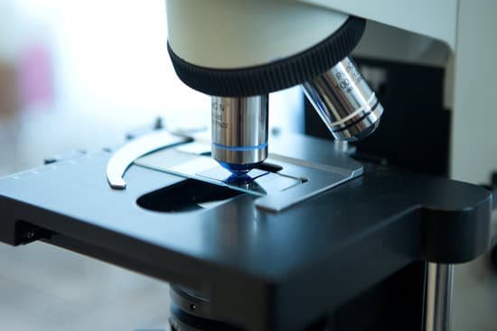
The stage of a microscope is the aluminum or iron platform where the specimen, usually on a glass slide, is raised or lowered for observation under the microscope. Microscope stages will often include stage clips that will hold the slide in place while the stage is being adjusted up and down or side to side.
What does west nile virus look like under a microscope?
(2003), like other flaviviruses, West Nile virus is a small virus, with a single stranded, positive sense RNA genome comprising about 11,000 nucleotides wrapped in a nucleocapsid and surrounded by a lipid membrane. Under a microscope the virions appear roughly as spheres 40-65 nm in diameter.
Can bacteria be seen under a light microscope?
Generally speaking, it is theoretically and practically possible to see living and unstained bacteria with compound light microscopes, including those microscopes which are used for educational purposes in schools.
What eukaryotic cellular structures are visible under a microscope?
The cell wall, nucleus, vacuoles, mitochondria, endoplasmic reticulum, Golgi apparatus, and ribosomes are easily visible in this transmission electron micrograph.
What is the greatest advantage of an electron microscope?
Electron microscopes have two key advantages when compared to light microscopes: They have a much higher range of magnification (can detect smaller structures) They have a much higher resolution (can provide clearer and more detailed images)
Which electron microscope sees through the specimen?
This technique allows you to see the surface of just about any sample, from industrial metals to geological samples to biological specimens like spores, insects, and cells.
How to view microscope on computer?
If the viewer is using the microscope with a computer, they may need to begin by loading the device’s software. Plug the device into any open USB port on the computer or the television. Hold the microscope and lightly touch the lens to the specimen. The image should now be visible on the monitor or television screen.
What is the most advanced microscope?
Lawrence Berkeley National Labs just turned on a $27 million electron microscope. Its ability to make images to a resolution of half the width of a hydrogen atom makes it the most powerful microscope in the world.
What is the advantage of using an electron microscope?
Electron microscopes have two key advantages when compared to light microscopes: They have a much higher range of magnification (can detect smaller structures) They have a much higher resolution (can provide clearer and more detailed images)
Does electron microscopes use permanent magnets?
Summary: A portable scanning electron microscope (SEM) column design is presented which makes use of permanent magnets. … The column is designed to be mod- ular, so that it can fit onto a wide range of different specimen chamber types, and can also be readily replaced.
What is ftir microscope?
Fourier transform infrared (FTIR) microscopes are used in conjunction with FTIR spectroscopy to allow visualization of a sample as an analysis of its components is being done. As the sample goes through the FTIR spectroscopy, it can be visualized using the microscope inside the spectrometer. …
Which microscope has the highest magnification power?
Out of all types of microscopes, the electron microscope has the greatest capability in achieving high magnification and resolution levels, enabling us to look at things right down to each individual atom.
Can you see atoms with a light microscope?
The size of a typical atom is about 10-10 m, which is 10,000 times smaller than the wavelength of light. Since an atom is so much smaller than the wavelength of visible light, it’s much too small to change the way light is reflected, so observing an atom with an optical microscope will not work.
How are electron microscopes used in our lives?
In life sciences, electron microscopes are being used to explore the molecular mechanisms of disease, to visualize the 3D architecture of tissues and cells, to unambiguously determine the conformation of flexible protein structures and complexes, and to observe individual viruses and macromolecular complexes in their …
How to take a picture through a microscope with iphone?
Open the application and focus the object correctly in the microscope. Bring the camera in the phone near the eye piece and click a photo once you get the object correctly focused. Hit ‘Use’ and put in the magnification of the image. Hit ‘Accept’ and view the image.
Who discovered the first microscope?
The development of the microscope allowed scientists to make new insights into the body and disease. It’s not clear who invented the first microscope, but the Dutch spectacle maker Zacharias Janssen (b. 1585) is credited with making one of the earliest compound microscopes (ones that used two lenses) around 1600.
When did microscopes invented?
Lens Crafters Circa 1590: Invention of the Microscope. Every major field of science has benefited from the use of some form of microscope, an invention that dates back to the late 16th century and a modest Dutch eyeglass maker named Zacharias Janssen.
What is the function of iris in microscope?
Iris Diaphragm controls the amount of light reaching the specimen. It is located above the condenser and below the stage. Most high quality microscopes include an Abbe condenser with an iris diaphragm. Combined, they control both the focus and quantity of light applied to the specimen.

