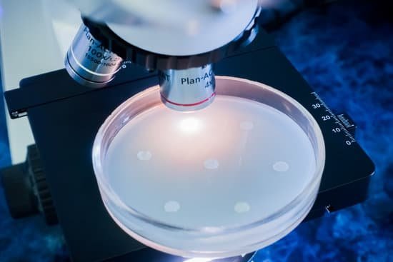
How to identify epithelial tissue under microscope? There are three basic shapes used to classify epithelial cells. A squamous epithelial cell looks flat under a microscope. A cuboidal epithelial cell looks close to a square. A columnar epithelial cell looks like a column or a tall rectangle.
How do you identify epithelial tissue? Epithelial tissues are identified by both the number of layers and the shape of the cells in the upper layers. There are eight basic types of epithelium: six of them are identified based on both the number of cells and their shape; two of them are named by the type of cell (squamous) found in them.
When looking under the microscope where should you look for epithelial tissue? 1. In observing epithelial cells under a microscope, the cells are arranged in a single layer and look tall and narrow, and the nucleus is located close to the basal side of the cell.
What does epithelial tissue look like? Epithelial tissue is scutoid shaped, tightly packed and forms a continuous sheet. It has almost no intercellular spaces. All epithelia is usually separated from underlying tissues by an extracellular fibrous basement membrane. The lining of the mouth, lung alveoli and kidney tubules are all made of epithelial tissue.
How to identify epithelial tissue under microscope? – Related Questions
Why are things flipped under a microscope?
Under the slide on which the object is being magnified, there is a light source that shines up and helps you to see the object better. This light is then refracted, or bent around the lens. Once it comes out of the other side, the two rays converge to make an enlarged and inverted image.
Why is the microscope made with an inclination joint?
Inclination Joint: Where the microscope arm connects to the microscope base, there may be a pin. If so, you can place one hand on the base and with the other hand grab the arm and rotate it back. It will tilt your microscope back for more comfortable viewing.
How to use a micrometer with a microscope?
Procedure. Place a stage micrometer on the microscope stage, and using the lowest magnification (4X), focus on the grid of the stage micrometer. Rotate the ocular micrometer by turning the appropriate eyepiece. Move the stage until you superimpose the lines of the ocular micrometer upon those of the stage micrometer.
What is electron microscope and how does it work?
The electron microscope uses a beam of electrons and their wave-like characteristics to magnify an object’s image, unlike the optical microscope that uses visible light to magnify images.
How to increase magnification of compound microscope?
From the above formula, we can conclude that the magnifying power of the compound microscope increases when the focal lengths of both objective and eyepiece lenses decrease.
When was the digital microscope invented?
The first digital Microscope was manufactured in 1986 in Tokyo, Japan which constituted a control box and a lens the was connected to the camera. This is currently known as the Hirox Co. LTD.
What is macroscopic and microscopic?
The macroscopic level includes anything seen with the naked eye and the microscopic level includes atoms and molecules, things not seen with the naked eye. Both levels describe matter. Matter is anything that occupies space and has mass and can be in three states: Solid, Liquid, or Gas.
When focusing a microscope which knob is used first?
When focusing on a slide, ALWAYS start with either the 4X or 10X objective. Once you have the object in focus, then switch to the next higher power objective. Re-focus on the image and then switch to the next highest power.
How to get rid of microscopic blood in urine?
Depending on the condition causing your hematuria, treatment might involve taking antibiotics to clear a urinary tract infection, trying a prescription medication to shrink an enlarged prostate or having shock wave therapy to break up bladder or kidney stones. In some cases, no treatment is necessary.
Which microscope provides a right side up image?
Which microscope provides a right-side-up image? In the design of a polarizing microscope, the polarizer is placed between the: light source and the sample stage.
What organelles can be seen with a compound microscope?
In most plant cells, the organelles that are visible under a compound light microscope are the cell wall, cell membrane, cytoplasm, central vacuole, and nucleus.
How do detectives use microscopes?
Microscopy can be applied in the identification of trace evidence such as fragments, fibers, hairs, fingerprints which are left the crime scene, on a victim or suspect. … Microscopes can be used to compare shattered glass left at the scene to that found on a suspect.
What limits the magnifying power of a microscope?
The limitations on resolution (and therefore magnifying power) imposed by the constraints of a simple microscope can be overcome by the use of a compound microscope, in which the image is relayed by two lens arrays. One of them, the objective, has a short focal length and is placed close to the object being examined.
How is the magnification of a microscope calculated?
The total magnification of the microscope is calculated from the magnifying power of the objective multiplied by the magnification of the eyepiece and, where applicable, multiplied by intermediate magnifications. … If an object is viewed with the eye from a distance of 250 mm, the magnification is 1x.
Can you see fluorescent dye with regular light microscope?
Using a ‘simple’ epifluorescence microscope, with a high NA objective, a sensitive camera, and a laser powerful enough to provide the required excitation intensity, you should be able to see individual fluorescent particles.
What to do when microscope lens needs to be cleaned?
Dip a lens wipe or cotton swab into distilled water and shake off any excess liquid. Then, wipe the lens using the spiral motion. This should remove all water-soluble dirt.
What is the angular aperture optical and optical microscope?
α is half of the angle of the light cone. The maximum longitudinal angle of the cone of light collected by the front lens of the objective is known as the ‘angular aperture’ (s. Figure 1). In addition to an increasing NA, image brightness is also proportional to the angular aperture.
What is the function of microscope?
A microscope is an instrument that can be used to observe small objects, even cells. The image of an object is magnified through at least one lens in the microscope. This lens bends light toward the eye and makes an object appear larger than it actually is.
What power microscope is needed to see blood cells?
At 400x magnification you will be able to see bacteria, blood cells and protozoans swimming around. At 1000x magnification you will be able to see these same items, but you will be able to see them even closer up.
What is a body tube used for on a microscope?
The microscope body tube separates the objective and the eyepiece and assures continuous alignment of the optics. It is a standardized length, anthropometrically related to the distance between the height of a bench or tabletop (on which the microscope stands) and the position of the seated observer’s…
Why is microscopic visualization not sufficient to properly identify bacteria?
Why is visualization not sufficient to properly identify bacteria? Bacteria have a limited set of shapes and many unrelated bacteria share the same shape. What is the hallmark of dichotomous keys?

