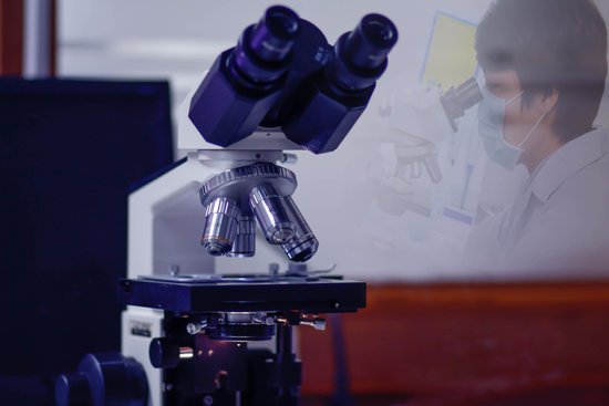
How to increase resolution in microscope? To achieve the maximum (theoretical) resolution in a microscope system, each of the optical components should be of the highest NA available (taking into consideration the angular aperture). In addition, using a shorter wavelength of light to view the specimen will increase the resolution.
How do you increase magnification and resolution? The only possibility to increase resolution is to switch to an objective with a higher resolving power, to use a shorter wavelength of light or to generally improve the optics. But there is a physical limit. A part of the above image was further magnified 2x.
How do you make a microscope image clearer? Turn the coarse focus knob slowly until you are able to see the cells. Turn the fine focus knob slowly until the cells are in focus and you can see them clearly. Repeat steps 1-5 using the higher power magnification to see the cells in more detail.
What two factors affect resolution of a microscope? As discussed above, the primary factor in determining resolution is the objective numerical aperture, but resolution is also dependent upon the type of specimen, coherence of illumination, degree of aberration correction, and other factors such as contrast enhancing methodology either in the optical system of the …
How to increase resolution in microscope? – Related Questions
How to create a scale bar for a microscope image?
In the ‘Analyze/Tools’ menu select ‘Scale Bar’. The scale bar dialog will open and a scale bar will appear on your image. You can adjust the size, color, and placement of your scale bar. Once you are finished click on ‘OK’, save your image, and you are done.
What does the resolving power of a microscope refer to?
Resolving power denotes the smallest detail that a microscope can resolve when imaging a specimen; it is a function of the design of the instrument and the properties of the light used in image formation. … The smaller the distance between the two points that can be distinguished, the higher the resolving power.
What you can do with a microscope?
A microscope is an instrument that can be used to observe small objects, even cells. The image of an object is magnified through at least one lens in the microscope. This lens bends light toward the eye and makes an object appear larger than it actually is.
What foods can you eat with microscopic colitis?
Avoid beverages that are high in sugar or sorbitol or contain alcohol or caffeine, such as coffee, tea and colas, which may aggravate your symptoms. Choose soft, easy-to-digest foods. These include applesauce, bananas, melons and rice. Avoid high-fiber foods such as beans and nuts, and eat only well-cooked vegetables.
What can you see under light microscope?
Explanation: You can see most bacteria and some organelles like mitochondria plus the human egg. You can not see the very smallest bacteria, viruses, macromolecules, ribosomes, proteins, and of course atoms.
When was electron microscopes invented?
Ernst Ruska, a German electrical engineer, is credited with inventing the electron microscope. The earliest electron microscope was developed in 1931, and the first commercial, mass-produced instrument became available in 1939.
What do the microscope and brush symbolize respectively?
What do the microscope and brush symbolize, respectively? The microscope symbolizes the author’s desires, and the brush symbolizes his inspiration.
Why is urine examined under a microscope?
This test looks at a sample of your urine under a microscope. It can see cells from your urinary tract, blood cells, crystals, bacteria, parasites, and cells from tumors. This test is often used to confirm the findings of other tests or add information to a diagnosis.
How to view a microscope on a computer?
If the viewer is using the microscope with a computer, they may need to begin by loading the device’s software. Plug the device into any open USB port on the computer or the television. Hold the microscope and lightly touch the lens to the specimen. The image should now be visible on the monitor or television screen.
How scanning tunneling microscope works?
The scanning tunneling microscope (STM) works by scanning a very sharp metal wire tip over a surface. By bringing the tip very close to the surface, and by applying an electrical voltage to the tip or sample, we can image the surface at an extremely small scale – down to resolving individual atoms.
What are the differences between light and electron microscopes?
Electron microscopes differ from light microscopes in that they produce an image of a specimen by using a beam of electrons rather than a beam of light. Electrons have much a shorter wavelength than visible light, and this allows electron microscopes to produce higher-resolution images than standard light microscopes.
How does the letter e look under a microscope?
The letter “e” appears upside down and backwards under a microscope. Either, diatoms are single celled, or they do not have a cell wall.
How would you carry a microscope?
Always carry the microscope with 2 hands—place one hand on the microscope arm and the other hand under the microscope base. Do not touch the objective lenses (i.e. the tips of the objectives). Keep the objectives in the scan position and keep the stage low when adding or removing slides.
What is the highest magnification your microscope can give?
Light microscopes combine the magnification of the eyepiece and an objective lens. Calculate the magnification by multiplying the eyepiece magnification (usually 10x) by the objective magnification (usually 4x, 10x or 40x). The maximum useful magnification of a light microscope is 1,500x.
What is the function of condenser lens in microscope?
On upright microscopes, the condenser is located beneath the stage and serves to gather wavefronts from the microscope light source and concentrate them into a cone of light that illuminates the specimen with uniform intensity over the entire viewfield.
How to look at sperm under microscope?
You can view sperm at 400x magnification. You do NOT want a microscope that advertises anything above 1000x, it is just empty magnification and is unnecessary. In order to examine semen with the microscope you will need depression slides, cover slips, and a biological microscope.
What are the 3 different types of microscopes?
There are three basic types of microscopes: optical, charged particle (electron and ion), and scanning probe. Optical microscopes are the ones most familiar to everyone from the high school science lab or the doctor’s office.
What does objective lens mean in microscope?
An objective lens is the most important optical unit that determines the basic performance/function of an optical microscope To provide an optical performance/function optimal for various needs and applications (i.e. the most important performance/function for an optical microscope), a wide variety of objective lenses …
What’s smaller than microscopic?
As adjectives the difference between microscopic and submicroscopic. is that microscopic is of, or relating to microscopes or microscopy; microscopal while submicroscopic is smaller than microscopic; too small to be seen even with a microscope.
What is the advantage of electron microscope?
Electron microscopes have two key advantages when compared to light microscopes: They have a much higher range of magnification (can detect smaller structures) They have a much higher resolution (can provide clearer and more detailed images)
Are convex lenses used in microscopes?
For the purposes of microscopy, convex lenses are used for their ability to focus light at a single point. … Microscopes borrowed this idea, using convex lenses to focus light towards a point that is f distance away from the lens. This distance is known as the focal length of the lens and depends on the shape.

