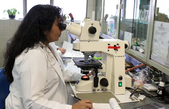
How to measure specimens with a microscope? The measurement of specimen size with a microscope is normally made by using an eyepiece graticule sometimes referred to as a reticule. This is a x10 eyepiece that has a scale inserted which is in focus at the same time as the specimen.
How is a microscope used to estimate the size of microscopic specimens? To estimate the size of an object seen with a microscope, first estimate what fraction of the diameter of the field of vision that the object occupies. Then multiply the diameter you calculated in micrometers by that fraction.
When should you use the coarse adjustment on a microscope? There are two knobs on the side of the arm that move the eyepiece. On some microscopes these are located closer to the eyepiece, on others they maybe closer to the stage. The larger knob is called the COARSE ADJUSTMENT and the smaller knob is the FINE ADJUSTMENT. The coarse adjustment is ONLY used on LOW power.
What does the coarse adjustment knob do quizlet? The coarse adjustment knob should only be used in the scanning objective lens because it moves the stage up and down in bigger increments and brings it closer to the lens faster, bringing it into focus.
How to measure specimens with a microscope? – Related Questions
What is a dental operating microscope?
Dental Operating Microscopes, or dental surgical microscopes, are designed to provide ideal magnification of the area of the mouth being worked on while also allowing the clinician to maintain an ergonomic working position. … In addition to the basic microscope, there are also additional options available.
What is the magnification of x ray microscopes?
1). Influencing the direction of the X-rays is not needed in the basic version of this type of microscope. The magnification of the set-up is the quotient L2 / L1 with the distance source-sample L1 and the distance source-image L2.
What determines resolution of a microscope?
In microscopy, the term ‘resolution’ is used to describe the ability of a microscope to distinguish detail. … The resolution of a microscope is intrinsically linked to the numerical aperture (NA) of the optical components as well as the wavelength of light which is used to examine a specimen.
What type of image does a tem microscope produce?
A Transmission Electron Microscope produces a high-resolution, black and white image from the interaction that takes place between prepared samples and energetic electrons in the vacuum chamber. Air needs to be pumped out of the vacuum chamber, creating a space where electrons are able to move.
Who made up the microscopes?
A Dutch father-son team named Hans and Zacharias Janssen invented the first so-called compound microscope in the late 16th century when they discovered that, if they put a lens at the top and bottom of a tube and looked through it, objects on the other end became magnified.
What is microscopic ear surgery?
This is also called as tympanic membrane perforation. Depending on your condition your Doctor may Recommend a Microscopic Ear Surgery. One such Surgery is called- Tympanoplasty. Tympanoplasty is the surgical operation that is performed to reconstruct the eardrum or the small bones of the middle ear.
How much money does an electron microscope cost?
The price of a new electron microscope can range from $80,000 to $10,000,000 depending on certain configurations, customizations, components, and resolution, but the average cost of an electron microscope is $294,000. The price of electron microscopes can also vary by type of electron microscope.
What is a convex lens of microscope?
A convex lens is thicker at the center than the edge and will focus a beam of light to a point a certain distance in front of the lens (the focal length). A concave lens is the opposite, being thicker at the edge than the center and spreading out a beam of light. Microscopes use convex lenses in order to focus light.
Are you see when you look in the microscope?
A microscope lets you look at and study very tiny things in great detail, which the naked eye cannot see. Even under a low-power optical microscope, the fine structures of specimens, or the objects under view, can be seen. … A photograph of the magnified view through a microscope is called a micrograph.
How to focus a simple microscope?
To focus a microscope, rotate to the lowest-power objective, and place your sample under the stage clips. Play with the magnification using the coarse adjustment knob and move your slide around until it is centered.
How to switch objectives on a microscope?
When focusing on a slide, ALWAYS start with either the 4X or 10X objective. Once you have the object in focus, then switch to the next higher power objective. Re-focus on the image and then switch to the next highest power.
What are the units used to measure microscopic objects?
micrometre. micrometre, also called micron, metric unit of measure for length equal to 0.001 mm, or about 0.000039 inch. Its symbol is μm. The micrometre is commonly employed to measure the thickness or diameter of microscopic objects, such as microorganisms and colloidal particles.
What does a ribosome look like under a microscope?
Ribosomes – which cannot be seen with a light microscope, but only using an electron microscope – are the site of protein synthesis. Even at very high magnification, they look like (pairs of) dots.
Can you see arterioles without microscope?
The macrovasculature is composed of those blood vessels that can be seen with the naked eye. The microvasculature is composed of blood vessels that are smaller than 100 microns may only be seen through the microscope.
What is a compound microscope in biology?
A compound microscope is a microscope that uses multiple lenses to enlarge the image of a sample. … Compound microscopes usually include exchangeable objective lenses with different magnifications (e.g 4x, 10x, 40x and 60x), mounted on a turret, to adjust the magnification.
Can small kidney stones cause microscopic blood in urine?
The stones are generally painless, so you probably won’t know you have them unless they cause a blockage or are being passed. Then there’s usually no mistaking the symptoms — kidney stones, especially, can cause excruciating pain. Bladder or kidney stones can also cause both gross and microscopic bleeding.
What kind of microscope can see genes?
To view the DNA as well as a variety of other protein molecules, an electron microscope is used. Whereas the typical light microscope is only limited to a resolution of about 0.25um, the electron microscope is capable of resolutions of about 0.2 nanometers, which makes it possible to view smaller molecules.
Does higher resolution mean things can be closer together microscope?
If the two points are closer together than your resolution then they will appear ill-defined and their positions will be inexact. A microscope may offer high magnification, but if the lenses are of poor quality the resulting poor resolution will degrade the image quality.
How was the first compound microscope different from leeuwenhoek& 39?
Whereas van Leeuwenhoek used a simple microscope, in which light is passed through just one lens, Galileo’s compound microscope was more sophisticated, passing light through two sets of lenses.
What is the function of objective lenses in a microscope?
The objective, located closest to the object, relays a real image of the object to the eyepiece. This part of the microscope is needed to produce the base magnification. The eyepiece, located closest to the eye or sensor, projects and magnifies this real image and yields a virtual image of the object.

