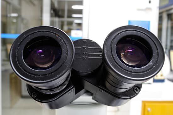
How to measure using a dissecting microscope? Multiply the magnification on the eyepiece (10x) by any magnification present on the nose piece (usually 1x, but it can be more) by the number on the magnification knob to get your total magnification.
How do you calculate the size of a cell using a microscope? Divide the number of cells in view with the diameter of the field of view to figure the estimated length of the cell. If the number of cells is 50 and the diameter you are observing is 5 millimeters in length, then one cell is 0.1 millimeter long. Measured in microns, the cell would be 1,000 microns in length.
What can you see with a dissecting microscope? A dissecting microscope is used to view three-dimensional objects and larger specimens, with a maximum magnification of 100x. This type of microscope might be used to study external features on an object or to examine structures not easily mounted onto flat slides.
What do electron microscopes allow you to see? Electron microscopy uses electrons to “see” small objects in the same way that light beams let us observe our surroundings or objects in a light microscope. With EM, we can look at the feather-like scales of an insect, the internal structures of a cell, individual proteins or even individual atoms in a metal alloy.
How to measure using a dissecting microscope? – Related Questions
What is microscope objectives?
The microscope objective is a key component for reaching high performance of a microscope. It is the part which is placed next to the observed object, usually in a fairly small distance of a few millimeters.
How to use a micrometer on a microscope?
Procedure. Place a stage micrometer on the microscope stage, and using the lowest magnification (4X), focus on the grid of the stage micrometer. Rotate the ocular micrometer by turning the appropriate eyepiece. Move the stage until you superimpose the lines of the ocular micrometer upon those of the stage micrometer.
What type of microscope uses a vacuum?
Most electron microscopes are high-vacuum instruments. Vacuums are needed to prevent electrical discharge in the gun assembly (arcing), and to allow the electrons to travel within the instrument unimpeded.
What does a compound light microscope use?
With higher levels of magnification than stereo microscopes, a compound microscope uses a compound lens to view specimens which cannot be seen at lower magnification, such as cell structures, blood, or water organisms.
How are light microscopes and electron microscopes the same?
Electron microscopes differ from light microscopes in that they produce an image of a specimen by using a beam of electrons rather than a beam of light. Electrons have much a shorter wavelength than visible light, and this allows electron microscopes to produce higher-resolution images than standard light microscopes.
What did the electron microscope discovered?
In 1931, two German scientists, Ernst Ruska and Max Knoll, found a way to achieve a resolution greater than that of light. They stopped using light, instead realizing they could transmit electrons through a specimen to form an image.
Is a light microscope or electron microscope better?
Electrons have much a shorter wavelength than visible light, and this allows electron microscopes to produce higher-resolution images than standard light microscopes.
How much does it cost to buy an electron microscope?
The price of a new electron microscope can range from $80,000 to $10,000,000 depending on certain configurations, customizations, components, and resolution, but the average cost of an electron microscope is $294,000. The price of electron microscopes can also vary by type of electron microscope.
Can you see the nucleus under a light microscope?
Thus, light microscopes allow one to visualize cells and their larger components such as nuclei, nucleoli, secretory granules, lysosomes, and large mitochondria. The electron microscope is necessary to see smaller organelles like ribosomes, macromolecular assemblies, and macromolecules.
What does microscopic blood in urine indicate?
Microscopic urinary bleeding is a common symptom of glomerulonephritis, an inflammation of the kidneys’ filtering system. Glomerulonephritis may be part of a systemic disease, such as diabetes, or it can occur on its own.
What is the function of the objective in a microscope?
The objective, located closest to the object, relays a real image of the object to the eyepiece. This part of the microscope is needed to produce the base magnification. The eyepiece, located closest to the eye or sensor, projects and magnifies this real image and yields a virtual image of the object.
How does a optical microscope works?
Light from a mirror is reflected up through the specimen, or object to be viewed, into the powerful objective lens, which produces the first magnification. The image produced by the objective lens is then magnified again by the eyepiece lens, which acts as a simple magnifying glass.
How to turn on light microscope?
Place your microscope on a flat surface and connect its power cord into an outlet. Now, flip on the light switch, which is typically located on the bottom of the microscope. After flipping the switch, the light should come out of the illuminator, which is the light source.
What does the 40x objective do on the microscope?
The high-powered objective lens (also called “high dry” lens) is ideal for observing fine details within a specimen sample.
How compound light microscope works?
A compound light microscope has its own light source in its base. The incandescent light from the light source is reflected by a condenser lens beneath the specimen, and the light passes through the specimen, up to the objective lens, then the projector lens sends the magnified image onto the eyepiece.
Where was the transmission electron microscope invented?
Ernst Ruska at the University of Berlin, along with Max Knoll, combined these characteristics and built the first transmission electron microscope (TEM) in 1931, for which Ruska was awarded the Nobel Prize for Physics in 1986.
Do light microscopes show color?
The magnified image that a light microscope produces contains color. In fact, if you use any ordinary optical microscope that magnifies up to 500x levels, then you’ll most likely see colors in the magnified image. … They produce grayscale images of the specimen, i.e., the magnified images are black and white.
Can cilia be seen with a light microscope?
Cilia, Microvilli and Stereocillia. Some apical specializations of epithelial cells are visible by light microscopy. Specifically when they are abundant. Due to their size, most cilia are easily recognizable.
How to connect microscope to computer?
Plug the device into any open USB port on the computer or the television. Hold the microscope and lightly touch the lens to the specimen. The image should now be visible on the monitor or television screen. These microscopes should only be used to examine dry specimens.
What part of the microscope changes the magnification?
Revolving Nosepiece or Turret: This is the part of the microscope that holds two or more objective lenses and can be rotated to easily change power (magnification).

