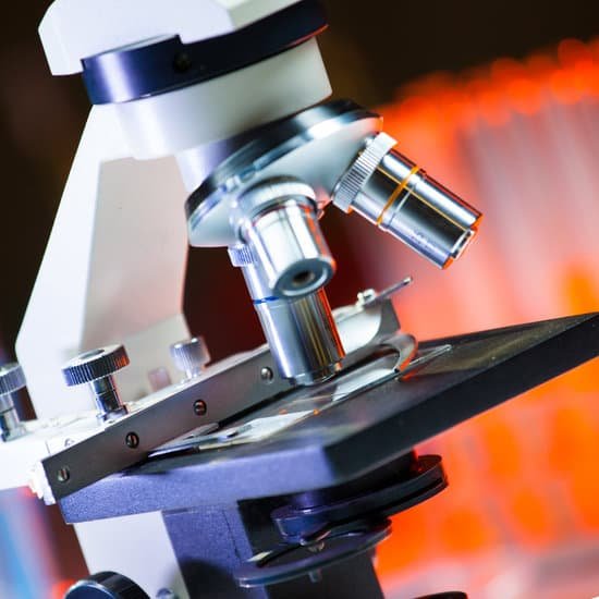
How to microscope sperm? To do a home test, a man would have to wait for around five minutes after ejaculation for the semen to liquefy, then apply a small amount to a plastic sheet and press it against the microscope for inspection. This can be done without getting semen on to the phone, says Kobori.
Can you see sperm in a microscope? The air-fixed, stained spermatozoa are observed under a bright-light microscope at 400x or 1000x magnification. Their viability and mor- phology can be analysed at the same time. Those appearing red-pink in colour have a damaged membrane whereas white sperm are viable, as in Photo 2.
Can you see sperm without a microscope? Better have a microscope, because sperm are far too tiny to see with the naked eye. How tiny? Each one measures about 0.002 inch from head to tail, or about 50 micrometers.
What microscope is used to see very small objects? Viewable objects by magnification.
How to microscope sperm? – Related Questions
What is a condenser knob on a microscope?
Condenser Focusing Knob – This control is used to precisely adjust the vertical height of the condenser. Condenser Lens – This lens system is located immediately under the stage and focuses the light on the specimen.
Can cystitis cause microscopic hematuria?
Microscopic hematuria is found in about half of cystitis cases; when found without symptoms or pyuria, it should prompt a search for malignancy. Other possibilities to be considered in the differential diagnosis include calculi, vasculitis, renal tuberculosis, and glomerulonephritis.
How many sets of lenses does a compound microscope have?
Typically, a compound microscope is used for viewing samples at high magnification (40 – 1000x), which is achieved by the combined effect of two sets of lenses: the ocular lens (in the eyepiece) and the objective lenses (close to the sample).
What’s the difference between a compound and a simple microscope?
A magnifying instrument that uses two types of lens to magnify an object with different zoom levels of magnification is called a compound microscope. … A magnifying instrument that uses only one lens to magnify objects is called a Simple microscope.
How does one properly carry a microscope?
The proper way to carry a microscope is to grasp the arm of the microscope with your dominant hand, lift the microscope up slowly, and use your other hand to firmly hold the base to stabilize the microscope as you transport it from one place to another.
How do you look at sperm under a microscope?
You can view sperm at 400x magnification. You do NOT want a microscope that advertises anything above 1000x, it is just empty magnification and is unnecessary. In order to examine semen with the microscope you will need depression slides, cover slips, and a biological microscope.
How to calculate microscope field of view?
For instance, if your eyepiece reads 10X/22, and the magnification of your objective lens is 40. First, multiply 10 and 40 to get 400. Then divide 22 by 400 to get a FOV diameter of 0.055 millimeters.
What type of evidence is viewed with a compound microscope?
Compound microscopes allow scientists to see microorganisms and cells. These microscopes are common today in science classrooms as well as laboratories.
Why is the light microscope a compound microscope?
The common light microscope used in the laboratory is called a compound microscope because it contains two types of lenses that function to magnify an object. The lens closest to the eye is called the ocular, while the lens closest to the object is called the objective.
Who was the first person to make a microscope?
A Dutch father-son team named Hans and Zacharias Janssen invented the first so-called compound microscope in the late 16th century when they discovered that, if they put a lens at the top and bottom of a tube and looked through it, objects on the other end became magnified.
What kind of microscopes are used in school?
The most common types of microscopes used in teaching are monocular light microscopes (80%), followed by binocular optical microscopes (16%), digital microscopes (3%), and stereomicroscopes (1%). A total of 43% of teachers perform microscopy using the demonstration method, and 37% of teachers use practical work.
Why should you carry a microscope with two hands?
Always carry the microscope with both hands. One hand should support the bottom, and the other should have a firm grip on the arm. … This will save energy, protect the specimens, and improve the longevity of the microscope. It will also keep the light from getting too hot to the touch.
How many objectives are on the microscope?
A typical microscope has three or four objective lenses with different magnifications, screwed into a circular “nosepiece” which may be rotated to select the required lens. These lenses are often color coded for easier use.
How do cancer cells look under a microscope?
Typically, the nucleus of a cancer cell is larger and darker than that of a normal cell and its size can vary greatly. Another feature of the nucleus of a cancer cell is that after being stained with certain dyes, it looks darker when seen under a microscope.
Can you see trichomoniasis under microscope?
Trichomoniasis can be diagnosed by looking at a sample of vaginal fluid for women or urine for men under a microscope. If the parasite can be seen under the microscope, no further tests are needed. If this test isn’t conclusive, tests called rapid antigen tests and nucleic acid amplification may be used.
How to help microscopic colitis?
Microscopic colitis can get better on its own, but most patients have recurrent symptoms. The main treatment for microscopic colitis is medication. In many cases, the doctor will start treatment with an antidiarrheal medication such as Pepto-Bismol® or Imodium® .
How to appropriately carry a microscope?
The proper way to carry a microscope is to grasp the arm of the microscope with your dominant hand, lift the microscope up slowly, and use your other hand to firmly hold the base to stabilize the microscope as you transport it from one place to another.
How does a microscope with reticle work?
Reticles are clear circular glass inserts with a scale inscribed on them. They sit right at the focal plane inside the eyepiece lens of the microscope and allow the investigator to make accurate measurements of specimens. In a stereo or binocular microscope, there will only be one reticle in one of the lenses.
Are microscopes like telescopes?
Although both instruments magnify objects so that the human eye can see them, a microscope looks at things very near, while telescopes view things very far away. This difference in purpose explains the substantial differences in their design.
How does a raman microscope work?
Raman spectroscopy works by shining a monochromatic light source—usually a laser—onto a sample and detecting the scattered light. … Plotting the intensity of the shifted light against the frequency produces a Raman spectrum of the sample.

