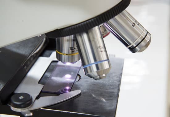
How to read magnification on microscope lenses? Microscope objective lenses will often have four numbers engraved on the barrel in a 2×2 array. The upper left number is the magnification factor of the objective. For example, 4x, 10x, 40x, and 100x. The upper right number is the numerical aperture of the objective.
How do you read the magnification of a microscope? To calculate the total magnification of the compound light microscope multiply the magnification power of the ocular lens by the power of the objective lens. For instance, a 10x ocular and a 40x objective would have a 400x total magnification. The highest total magnification for a compound light microscope is 1000x.
What is the total magnification at 4x 10x and 40x?
What does 40x on a microscope mean? It’s easy to understand. A 40x objective makes things appear 40 times larger than they actually are. … The product of the objective magnification and the eyepiece magnification gives the final magnification of the microscope. So, a 60x objective and a 10x eyepiece gives a total magnification of 600x.
How to read magnification on microscope lenses? – Related Questions
Are some fleas microscopic?
Fleas are not microscopic, they’re small but they can be seen with the naked eye. … The adult cat flea (the main flea on pets) is about 1/8 inch long and will be burrowed down at the base of the hairs on your dog.
What is the highest magnification of electron microscope?
The resolution limit of electron microscopes is about 0.2nm, the maximum useful magnification an electron microscope can provide is about 1,000,000x.
Why can light microscopes produce images in their natural color?
The magnified image that a light microscope produces contains color. … This is because in order to see something under a microscope, the object must have a very thin cross-section. In addition to that, it also needs to be thin enough for light to pass through it (generally).
What cells can you not see with a light microscope?
Some cell parts, including ribosomes, the endoplasmic reticulum, lysosomes, centrioles, and Golgi bodies, cannot be seen with light microscopes because these microscopes cannot achieve a magnification high enough to see these relatively tiny organelles.
Where was the microscope made?
Lippershey settled in Middelburg, where he made spectacles, binoculars and some of the earliest microscopes and telescopes. Also living in Middelburg were Hans and Zacharias Janssen. Historians attribute the invention of the microscope to the Janssens, thanks to letters by the Dutch diplomat William Boreel.
How do you properly use a compound light microscope?
Turn the revolving turret (2) so that the lowest power objective lens (eg. 4x) is clicked into position. Place the microscope slide on the stage (6) and fasten it with the stage clips. Look at the objective lens (3) and the stage from the side and turn the focus knob (4) so the stage moves upward.
What is a revolving turret on a microscope?
Revolving Nosepiece or Turret: This is the part that holds two or more objective lenses and can be rotated to easily change power. Objective Lenses: Usually you will find 3 or 4 objective lenses on a microscope. They almost always consist of 4X, 10X, 40X and 100X powers.
How doctors use microscopes?
Without microscopes, several diseases and illnesses can’t be identified, particularly cellular diseases. … By examining samples using such sensitive microscopes, doctors can accurately diagnose the types of microorganisms living in your body or the levels of certain kinds of cells in the body.
What did robert hooke discovered with a compound microscope?
While observing cork through his microscope, Hooke saw tiny boxlike cavities, which he illustrated and described as cells. He had discovered plant cells! Hooke’s discovery led to the understanding of cells as the smallest units of life—the foundation of cell theory.
What is the eyepiece number of a microscope?
For example, an eyepiece having a magnification of 10x typically has a field number ranging between 16 and 18 millimeters, while a lower magnification eyepiece (5x) has a field number of about 20 millimeters.
Why we put stain in microscope?
The most basic reason that cells are stained is to enhance visualization of the cell or certain cellular components under a microscope. Cells may also be stained to highlight metabolic processes or to differentiate between live and dead cells in a sample.
Can you see a cell in a microscope?
Microscopes provide magnification that allows people to see individual cells and single-celled organisms such as bacteria and other microorganisms. Types of cells that can be viewed under a basic compound microscope include cork cells, plant cells and even human cells scraped from the inside of the cheek.
What is refraction microscope?
The underlying principal of a microscope is that lenses refract light which allows for magnification. Refraction occurs when light travels through an area of space that has a changing index of refraction. … It is actually the water acting much like a lens in a microscope that gives it the appearance of bending.
What does smooth muscle look like under a microscope?
smooth muscle, also called involuntary muscle, muscle that shows no cross stripes under microscopic magnification. It consists of narrow spindle-shaped cells with a single, centrally located nucleus.
Who invented transmission electron microscope?
Ernst Ruska at the University of Berlin, along with Max Knoll, combined these characteristics and built the first transmission electron microscope (TEM) in 1931, for which Ruska was awarded the Nobel Prize for Physics in 1986.
What is used to move the slide on a microscope?
Stage clips hold the slides in place. If your microscope has a mechanical stage, the slide is controlled by turning two knobs instead of having to move it manually. One knob moves the slide left and right, the other moves it forward and backward.
What is the mechanical stage in a microscope?
The mechanical stage in a microscope is a mechanism that’s been mounted on the stage to hold the microscope slide in order to hold it steady and to reposition it when needed.
What is different about an electron microscope?
As the wavelength of an electron can be up to 100,000 times shorter than that of visible light photons, electron microscopes have a higher resolving power than light microscopes and can reveal the structure of smaller objects.
What structure on the microscope are the objectives attached to?
Nosepiece: The upper part of a compound microscope that holds the objective lens. Also called a revolving nosepiece or turret.
What does streptococcus pneumoniae look like under a microscope?
Streptococcus pneumoniae bacteria are gram-positive cocci arranged in chains and pairs (diplococci) on microscopic examination. A green, α-hemolytic, zone surrounds S. pneumoniae colonies on blood-agar plates.
How do the compound microscopes benefit us?
Compound microscopes allow scientists to see microorganisms and cells. … Without these microscopes, we would not know about the existence of cells and therefore would not be able to study DNA or make medical advances based on our knowledge of how different diseases or conditions attack cells.

