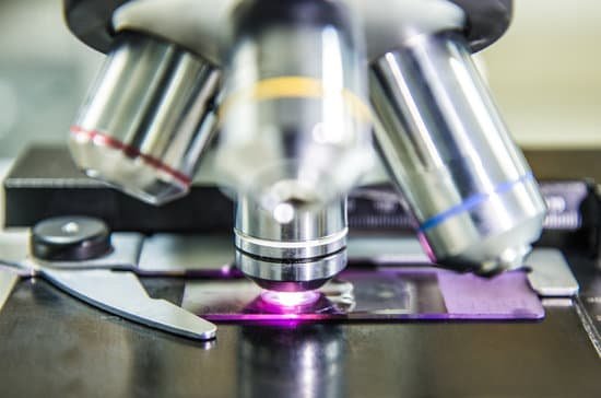
How to see tardigrades under microscope? To see tardigrades under the microscope, take your wet mount, and search for them, starting with the lowest power. You should be able to see one even at 40X total magnification.
Can you see tardigrades with a microscope? Yes, You Can See Tardigrades with a Cheap Optical Microscope.
How strong of a microscope Do you need to see tardigrades? In the right light you can actually see them with the naked eye. But researchers who work with tardigrades see them as they appear through a dissecting microscope of 20- to 30-power magnification—as charismatic miniature animals. Most tiny invertebrates dart about frantically.
How can I see a tardigrade? Many tardigrades are aquatic, but the easiest place for humans to find them is in damp moss, lichen, or leaf litter. Search in forests, around ponds, or even in your backyard. Your best bet is to look in damp places, where tardigrades are active.
How to see tardigrades under microscope? – Related Questions
What type of microscope is used in classrooms?
The most common types of microscopes used in teaching are monocular light microscopes (80%), followed by binocular optical microscopes (16%), digital microscopes (3%), and stereomicroscopes (1%). A total of 43% of teachers perform microscopy using the demonstration method, and 37% of teachers use practical work.
How is microscope made?
Lenses are given an antireflective coating, usually of magnesium fluoride. The eyepiece, the objective, and most of the hardware components are made of steel or steel and zinc alloys. A child’s microscope may have an external body shell made of plastic, but most microscopes have an body shell made of steel.
Who improved the microscope?
Galileo Galilei soon improved upon the compound microscope design in 1609. Galileo called his device an occhiolino, or “little eye.” English scientist Robert Hooke improved the microscope, too, and explored the structure of snowflakes, fleas, lice and plants.
Can some bacteria be seen without a microscope?
Yes. Most bacteria are too small to be seen without a microscope, but in 1999 scientists working off the coast of Namibia discovered a bacterium called Thiomargarita namibiensis (sulfur pearl of Namibia) whose individual cells can grow up to 0.75mm wide.
Why do light microscopes produce images in colour?
The magnified image that a light microscope produces contains color. … This is because in order to see something under a microscope, the object must have a very thin cross-section. In addition to that, it also needs to be thin enough for light to pass through it (generally).
What are plants made of at the microscopic scale?
At the microscopic scale, we can see that plants are made of cells, and that the cells have smaller parts such as cell walls, nuclei, and chloroplasts.
What did robert koch find using a compound microscope?
In the final decades of the 19th century, Koch conclusively established that a particular germ could cause a specific disease. He did this by experimentation with anthrax. Using a microscope, Koch examined the blood of cows that had died of anthrax. He observed rod-shaped bacteria and suspected they caused anthrax.
What does a nosepiece on a microscope?
Nosepiece houses the objectives. The objectives are exposed and are mounted on a rotating turret so that different objectives can be conveniently selected. Standard objectives include 4x, 10x, 40x and 100x although different power objectives are available. Coarse and Fine Focus knobs are used to focus the microscope.
What is simple microscope in physics?
A simple microscope is a magnifying glass that has a double convex lens with a short focal length. The examples of this kind of instrument include the hand lens and reading lens. When an object is kept near the lens, then its principal focus with an image is produced, which is erect and bigger than the original object.
Can electron microscopes see molecules?
That’s what it’s been like trying to take a picture of a molecule. … Advanced electron microscopes can get amazing resolution, fine enough to see inside an atom, but molecular bonds usually aren’t strong enough to hold up to their scrutiny.
Did galileo invent microscope?
In the late 16th century several Dutch lens makers designed devices that magnified objects, but in 1609 Galileo Galilei perfected the first device known as a microscope. Dutch spectacle makers Zaccharias Janssen and Hans Lipperhey are noted as the first men to develop the concept of the compound microscope.
Which microscope has the best magnification and resolution?
Out of all types of microscopes, the electron microscope has the greatest capability in achieving high magnification and resolution levels, enabling us to look at things right down to each individual atom.
Which object is used to focus light in a microscope?
A good quality microscope has a built-in illuminator, adjustable condenser with aperture diaphragm (contrast) control, mechanical stage, and binocular eyepiece tube. The condenser is used to focus light on the specimen through an opening in the stage.
How to look at your sperm thru a microscope?
You can view sperm at 400x magnification. You do NOT want a microscope that advertises anything above 1000x, it is just empty magnification and is unnecessary. In order to examine semen with the microscope you will need depression slides, cover slips, and a biological microscope.
What is the magnification of a scanning electron microscope?
An SEM can magnify a sample by about one million times (1,000,000x) at the most. Because a sample can be used in its natural state, the SEM is the easiest electron microscope to use.
What are the glass things in a microscope do?
A microscope slide is a thin flat piece of glass, typically 75 by 26 mm (3 by 1 inches) and about 1 mm thick, used to hold objects for examination under a microscope. Typically the object is mounted (secured) on the slide, and then both are inserted together in the microscope for viewing.
What scientist studies microscopic organisms?
Microbiologists study the microscopic organisms that cause infections, including viruses, bacteria, fungi and algae. They focus on the identification and growth of these organisms in order to understand their characteristics, with the overall aim to prevent, diagnose and treat infectious diseases.
Why is it convenient to have a parfocal microscope?
Why is it convenient to have a parfocal microscope? Parfocal microscopes stays in focus while the magnification is being changed.
How to calculate numerical aperture of microscope?
The highest angular aperture obtainable with a standard microscope objective would theoretically be 180 degrees, resulting in a value of 90 degrees for the half-angle used in the numerical aperture equation.
What do electrons travel through in an electron microscope?
An electron gun at the top of a TEM emits electrons that travel through the microscope’s vacuum tube. Rather than having a glass lens focusing the light (as in the case of light microscopes), the TEM employs an electromagnetic lens which focuses the electrons into a very fine beam.
Can u see bacteria without a microscope?
Yes. Most bacteria are too small to be seen without a microscope, but in 1999 scientists working off the coast of Namibia discovered a bacterium called Thiomargarita namibiensis (sulfur pearl of Namibia) whose individual cells can grow up to 0.75mm wide.

