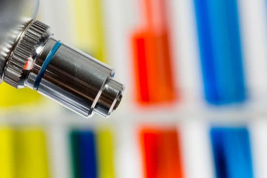
How to set up a compound light microscope? Turn the revolving turret (2) so that the lowest power objective lens (eg. 4x) is clicked into position. Place the microscope slide on the stage (6) and fasten it with the stage clips. Look at the objective lens (3) and the stage from the side and turn the focus knob (4) so the stage moves upward.
What is the correct order in which light passes through a compound microscope? The path of light begins with the illuminator, then passes through the condenser, the specimen, the objective lens, then then the ocular lens.
What are the proper ways of using compound microscope? When using a compound microscope, the correct viewing technique is to keep both eyes open. Look through the eyepiece with one eye, and look outside the microscope with the other eye. When you use the 10x lens to magnify the image, it may help to lower the amount of light for better clarity.
How is depth of focus determined? The essential distinction between the terms is clear: depth of field refers to object space and depth of focus to image space. A possibly useful mnemonic is that the field of view is that part of the object that is being examined, and the focus is the point at which parallel rays converge after passing through a lens.
How to set up a compound light microscope? – Related Questions
How many types microscopes are there?
There are several different types of microscopes used in light microscopy, and the four most popular types are Compound, Stereo, Digital and the Pocket or handheld microscopes. Some types are best suited for biological applications, where others are best for classroom or personal hobby use.
Why was the discovery of the microscope important?
The invention of the microscope allowed scientists to see cells, bacteria, and many other structures that are too small to be seen with the unaided eye. It gave them a direct view into the unseen world of the extremely tiny.
Can atoms be seen with electron microscope?
“So we can regularly see single atoms and atomic columns.” That’s because electron microscopes use a beam of electrons rather than photons, as you’d find in a regular light microscope. As electrons have a much shorter wavelength than photons, you can get much greater magnification and better resolution.
Did robert hooke made the first microscope?
Although Hooke did not make his own microscopes, he was heavily involved with the overall design and optical characteristics. The microscopes were actually made by London instrument maker Christopher Cock, who enjoyed a great deal of success due to the popularity of this microscope design and Hooke’s book.
What should you use to clean the microscope lens?
Dip a lens wipe or cotton swab into distilled water and shake off any excess liquid. Then, wipe the lens using the spiral motion. This should remove all water-soluble dirt.
Who invented the microscope and why?
A Dutch father-son team named Hans and Zacharias Janssen invented the first so-called compound microscope in the late 16th century when they discovered that, if they put a lens at the top and bottom of a tube and looked through it, objects on the other end became magnified.
Qué son las algas microscópicas?
Las algas microscópicas son en su mayoría unicelulares, viven en medios acuáticos formando el fitoplancton. Realizan la mayor parte de la fotosíntesis de la Tierra, siendo el primer eslabón de las cadenas tróficas de los ecosistemas acuáticos, liberando grandes cantidades de oxígeno a la atmósfera.
Can you see molecular movement with a microscope?
Electron microscopes allow scientists to see the structure of microorganisms, cells, metals, crystals and other tiny structures that weren’t visible with light microscopes. …
Is it normal to have microscopic blood in urine?
While in many instances the cause is harmless, blood in urine (hematuria) can indicate a serious disorder. Blood that you can see is called gross hematuria. Urinary blood that’s visible only under a microscope (microscopic hematuria) is found when your doctor tests your urine.
Can a light microscope see bacteria?
Generally speaking, it is theoretically and practically possible to see living and unstained bacteria with compound light microscopes, including those microscopes which are used for educational purposes in schools.
How many objectives are on the virtual microscope?
The microscope turret can simulate five different objectives, with the current objective highlighted. Click on a new objective to change the current magnification.
What do optical microscopes use for illumination and focus?
The light microscope is an instrument for visualizing fine detail of an object. It does this by creating a magnified image through the use of a series of glass lenses, which first focus a beam of light onto or through an object, and convex objective lenses to enlarge the image formed.
How many objectives does your microscope have?
Objective Lenses: Usually you will find 3 or 4 objective lenses on a microscope. They almost always consist of 4x, 10x, 40x and 100x powers. When coupled with a 10x (most common) eyepiece lens, total magnification is 40x (4x times 10x), 100x , 400x and 1000x.
What is the function of a microscope slide cover?
When viewing any slide with a microscope, a small square or circle of thin glass called a coverslip is placed over the specimen. It protects the microscope and prevents the slide from drying out when it’s being examined. The coverslip is lowered gently onto the specimen using a mounted needle .
What can be observed with a scanning electron microscope?
The SEM is routinely used to generate high-resolution images of shapes of objects (SEI) and to show spatial variations in chemical compositions: 1) acquiring elemental maps or spot chemical analyses using EDS, 2)discrimination of phases based on mean atomic number (commonly related to relative density) using BSE, and 3 …
How to tell magnification on dissecting microscope?
To change magnifications with a dissecting microscope, simply turn the knob located on the side of the scope. Examine the magnification knob. Some dissecting scopes will have total magnification written on the magnification knob so that you will not need to do multiplication to determine it.
What does the scanner lens do on the microscope?
A scanning objective lens provides the lowest magnification power of all objective lenses. 4x is a common magnification for scanning objectives and, when combined with the magnification power of a 10x eyepiece lens, a 4x scanning objective lens gives a total magnification of 40x.
What are the six basic components of a compound microscope?
The microscope optical train typically consists of an illuminator (including the light source and collector lens), a substage condenser, specimen, objective, eyepiece, and detector, which is either some form of camera or the observer’s eye (Table 1).
What is the source of illumination in the electron microscope?
An electron microscope is a microscope that uses a beam of accelerated electrons as a source of illumination.
What is the magnification of a laser scanning confocal microscope?
This first-generation instrument images corneal structures at ×400 magnification and has a field of view of 400 × 400 µm when used with a ×63 objective lens that has a numerical aperture of 0.9. It uses a 670 nm red wavelength Helium-Neon diode laser as its illumination source.

