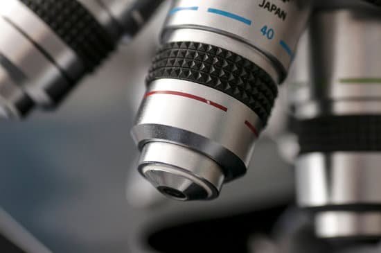
How to tell real stone from fake under microscope? To determine if your diamond is real, hold a magnifying glass up and look at the diamond through the glass. Look for imperfections within the stone. If you’re unable to find any, then the diamond is most likely fake. the majority of real diamonds have imperfections referred to as inclusions.
How do you tell if a diamond is real with a flashlight? A sparkle test is quick and easy to do since all you need are your eyes. Simply hold your diamond under a normal lamp and observe the bright shimmers of light bouncing off the diamond. A real diamond provides an exceptional sparkle since it reflects white light extremely well.
Does cubic zirconia float? Diamonds are dense and will sink quickly, while certain imitations will sink more slowly. If your gem doesn’t immediately sink to the bottom, it’s likely a glass or quartz imitation. However, other imitations, including cubic zirconia, will also sink quickly.
How can you tell if a stone is CZ? The best way to tell a cubic zirconia from a diamond is to look at the stones under natural light: a diamond gives off more white light (brilliance) while a cubic zirconia gives off a noticeable rainbow of colored light (excessive light dispersion).
How to tell real stone from fake under microscope? – Related Questions
What provides light on a microscope?
Illuminator is the light source for a microscope, typically located in the base of the microscope. … Iris Diaphragm controls the amount of light reaching the specimen. It is located above the condenser and below the stage. Most high quality microscopes include an Abbe condenser with an iris diaphragm.
What is the importance of a microscope in biology?
When it comes to biology, Microscopes are important because biology mainly deals with the study of cells (and their contents), genes and all organisms. Some organisms are so small that they can only be seen by using magnifications of 40x-1000x, which can only be achieved with the use of a microscope.
Who made the microscope?
The development of the microscope allowed scientists to make new insights into the body and disease. It’s not clear who invented the first microscope, but the Dutch spectacle maker Zacharias Janssen (b. 1585) is credited with making one of the earliest compound microscopes (ones that used two lenses) around 1600.
Do phase contrast microscopes use dyes?
In conclusion, contrast in phase imaging may be augmented by using dyes that increase the index of refraction, of which corroles are an example at 488-nm excitation. Quantitative phase imaging is therefore a technique amenable to specific labeling.
Why are images flipped in a microscope?
The eyepiece of the microscope contains a 10x magnifying lens, so the 10x objective lens actually magnifies 100 times and the 40x objective lens magnifies 400 times. There are also mirrors in the microscope, which cause images to appear upside down and backwards.
Is compound microscope stronger than an electron?
Because a compound microscope uses light, its resolution is limited to . 05 micrometer. … Electrons, however, have a much smaller wavelength, and therefore the total magnification of a scanning electron microscope is 200,000 times with a resolution of . 02 nanometer.
What does the coarse adjustment knob do on a microscope?
4. COARSE ADJUSTMENT KNOB — A rapid control which allows for quick focusing by moving the objective lens or stage up and down. It is used for initial focusing.
What is magnification of the microscope?
Magnification on a microscope refers to the amount or degree of visual enlargement of an observed object. Magnification is measured by multiples, such as 2x, 4x and 10x, indicating that the object is enlarged to twice as big, four times as big or 10 times as big, respectively.
What increases the magnification on a microscope?
A simple hand lens can increase the magnification and resolution by about 20 times their actual size by increasing the visual angle. A hand lens is often used to observe objects in the field. A compound microscope is capable of magnifying objects up to 1000 times their actual size.
How to identify malaria parasite under the microscope?
Malaria parasites can be identified by examining under the microscope a drop of the patient’s blood, spread out as a “blood smear” on a microscope slide. Prior to examination, the specimen is stained (most often with the Giemsa stain) to give the parasites a distinctive appearance.
What is tpm microscope?
TPM is a two photon-excited nonlinear fluorescence microscopy that enables the observation of deep tissues up to several hundred micrometers. … TPM enables observation of autofluorescence at the cellular level, and thus may provide new insights into the fluorescent molecules in/around RPE cells.
What cell organelles can be seen with an electron microscope?
The cell wall, nucleus, vacuoles, mitochondria, endoplasmic reticulum, Golgi apparatus, and ribosomes are easily visible in this transmission electron micrograph.
What does an electron microscope have vs a light microscope?
Electron microscopes differ from light microscopes in that they produce an image of a specimen by using a beam of electrons rather than a beam of light. Electrons have much a shorter wavelength than visible light, and this allows electron microscopes to produce higher-resolution images than standard light microscopes.
How do we clean the lens of microscope?
Dip a lens wipe or cotton swab into distilled water and shake off any excess liquid. Then, wipe the lens using the spiral motion. This should remove all water-soluble dirt.
What is a scanning electron microscope good for?
The SEM is routinely used to generate high-resolution images of shapes of objects (SEI) and to show spatial variations in chemical compositions: 1) acquiring elemental maps or spot chemical analyses using EDS, 2)discrimination of phases based on mean atomic number (commonly related to relative density) using BSE, and 3 …
What is a biological microscope used for?
A biological microscope is generally a type of optical microscope that is primarily designed to observe cells, tissues, and other biological specimens. Multiple objective lenses can be attached, which gives these microscopes a magnification that can range anywhere from 10x – 1,000x or more.
Can you see bacteria in a 10x zoom microscope?
While some eucaryotes, such as protozoa, algae and yeast, can be seen at magnifications of 200X-400X, most bacteria can only be seen with 1000X magnification. This requires a 100X oil immersion objective and 10X eyepieces.. Even with a microscope, bacteria cannot be seen easily unless they are stained.
How to clean a microscope at home?
Using a lint-free cotton swab, dip the end into your cleaning solution; alcohol,etc. Shake off excess fluid from the swab. Using the cotton end of the stick, start at the center of the lens using a circular motion and work your way to the outer edge. Gently wipe off any excess liquid with another dry lint-free swab.
What other differences a compound and dissecting microscopes?
Most importantly, dissecting microscopes are for viewing the surface features of a specimen, whereas compound microscopes are designed to look through a specimen.
How does the resolving power of a microscope change when?
(i) When the diameter of the objective lens is decreased β decreases so resolving power decreases. (ii) When the wavelength of the incident light is increased resolving power decreases.
How are microscopes used in the study of cells?
“to look at”) to study them. … This occurs because microscopes use two sets of lenses to magnify the image.

