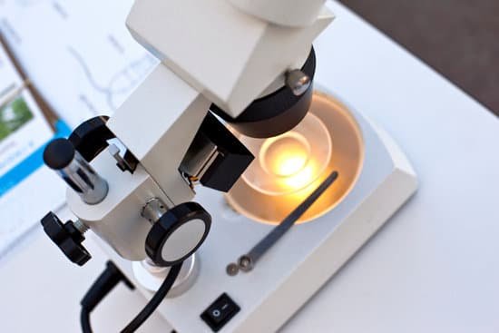
How to use a dark field microscope? To view a specimen in dark field, an opaque disc is placed underneath the condenser lens, so that only light that is scattered by objects on the slide can reach the eye. Instead of coming up through the specimen, the light is reflected by particles on the slide.
When would you use a dark field microscope? A dark field microscope is ideal for viewing objects that are unstained, transparent and absorb little or no light. These specimens often have similar refractive indices as their surroundings, making them hard to distinguish with other illumination techniques.
How do you use a field microscope? Darkfield is used to study marine organisms such as algae, plankton, diatoms, insects, fibers, hairs, yeast and protozoa as well as some minerals and crystals, thin polymers and some ceramics. Darkfield is used to study mounted cells and tissues.
Where is dark field microscope used? Darkfield illumination is a technique in optical microscopy that eliminates scattered light from the sample image. This yields an image with a dark background around the specimen, and is essentially the complete opposite of the brightfield illumination technique.
How to use a dark field microscope? – Related Questions
Can you see a snowflake without a microscope?
You can’t see the small particle that the water vapor crystallizes around with the naked eye or even a standard microscope.
Are microscopic pieces of plastic showing up in peoples blood?
17, 2020 (HealthDay News) — Microscopic bits of plastic have most likely taken up residence in all of the major filtering organs in your body, a new lab study suggests. Researchers found evidence of plastic contamination in tissue samples taken from the lungs, liver, spleen and kidneys of donated human cadavers.
What is an electron microscope lens made of?
Glass lenses of course, would impede electrons, therefore electron microscope (EM) lenses are electromagnetic converging lenses. A tightly wound wrapping of copper wire makes up the magnetic field that is the essence of the lens.
What is the mechanical stage control on a microscope?
A mechanical stage is a mechanism mounted on the stage that holds and moves the microscope slide. It has two knobs and allows the user to move the slide in the X or Y direction very smoothly and slowly by turning these knobs.
What strength microscope to see blood cells?
At 400x magnification you will be able to see bacteria, blood cells and protozoans swimming around. At 1000x magnification you will be able to see these same items, but you will be able to see them even closer up.
How discovered the microscope?
In the late 16th century several Dutch lens makers designed devices that magnified objects, but in 1609 Galileo Galilei perfected the first device known as a microscope.
Can you shade when drawing observations from a microscope?
You can shade using a number of hatching techniques but the ones that I have found most useful are: Stipple technique – using your reference drawing, begin to shade in the darker areas using a number of tiny dots.
What is the eyepiece magnification for a microscope?
The standard eyepiece magnifies 10x. Check the objective lens of the microscope to determine the magnification, which is usually printed on the casing of the objective.
What does ebola look like under a microscope?
The Ebola virus is different: it looks like a strand of spaghetti. And, if you look at an infected cell under an electron microscope, it looks like a ball of spaghetti coming out. Each virus is a long, flexible filament that can adopt different shapes.
What is the function of a microscope condenser?
On upright microscopes, the condenser is located beneath the stage and serves to gather wavefronts from the microscope light source and concentrate them into a cone of light that illuminates the specimen with uniform intensity over the entire viewfield.
What does a diaphragm lever do on a microscope?
Iris diaphragm lever- The iris diaphragm lever is the arm attached to the base of the condenser that regulates the amount of light passing through the condenser. The iris diaphragm permits the best possible contrast when vieweing the specimen.
Is a fluorescence microscope a compound microscope?
Most modern microscopes are compound microscopes, because the additional magnification gives a more enlarged image. … If only white light is used for illumination, then it’s bright-field microscopy. Figure 2.
What is the advantage of a dissecting microscope?
With a dissecting microscope whole objects can be viewed in three dimensions. Samples do not need to be sliced, and larger, live animals can be observed. Light can be passed through from underneath the sample, but also from the top or side using an external light source.
When was the microscope invented by robert hooke?
During this period, Hooke’s interest in microscopy and astronomy soared, and he published Micrographia, his best known work on optical microscopy in 1665. The next year, Hooke published a volume on comets, Cometa, detailing his close observation of the comets occurring in 1664 and 1665.
How to view blood under a microscope?
Place the slide on the microscope stage, and bring into focus on low power (100X). Adjust lighting and then switch into high power (400X). You should see hundreds of tiny red blood cells; there are billions circulating throughout our blood stream.
What did louis pasteur discovered with a compound microscope?
In the comound microscope, Louis Pasteur experimented with paratartaric acid and discovered that it did not rotate the plane angle of polarized light…
What microscope to use to do research in butterfly?
At present, the conventional observing and measuring method for the structure of butterfly wing uses high-power microscopies, such as scanning electron microscopy (SEM), transverse electron microscopy (TEM), scanning probe microscopy (SPM), scanning tunnel microscopy (STM), atomic force microscopy (AFM), etc.
How does the ion microscope work?
The helium ion microscope is a new type of microscope that uses helium ions for surface imaging and analysis. Its functionality is similar to a scanning electron microscope, but it uses a focused beam of helium ions instead of electrons. … SEM’s typically produce one secondary electron for each incoming electron.
How many types of microscope do we have?
There are several different types of microscopes used in light microscopy, and the four most popular types are Compound, Stereo, Digital and the Pocket or handheld microscopes. Some types are best suited for biological applications, where others are best for classroom or personal hobby use.
What’s the invincible microscopic animal?
What is a tardigrade? Tardigrades are microscopic eight-legged animals that have been to outer space and would likely survive the apocalypse.
What is the limit of resolution of a compound microscope?
The wavelength of visible light ranges from about 400 to 700 nanometers. The best compound microscopes cannot resolve parts of a specimen that are closer together than about 200 nanometers.

