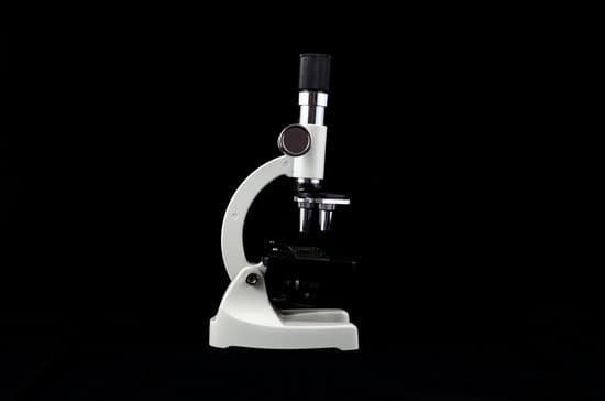
How was electron microscope invented? In 1931, Ruska built the first electron lens, an electromagnet that focused a beam of electrons just as an optical lens focuses a beam of light. His first electron microscope (1931) used two magnetic coils (electron lenses) in series.
Who invented electron microscope in 1935? The scanning electron microscope (SEM) was invented by Max Knoll in 1935, at the Telefunken Company in Berlin, for studying the secondary emission properties of television camera tube targets [1]; four years earlier, he and Ernst Ruska had built the first transmission electron microscope (TEM).
Who invented the electron microscope in 1940? 1940: Vladimir Zworykin, better known as a co-inventor of television, demonstrates the first electron microscope in the United States.
Why was the invention of the electron microscope so important? The electron microscope ushered in a new era of discoveries printed in academic journals. Atoms were seen by the human eye, as opposed to being merely conceived of. Knowledge of cell structures in plant and animal life increased dramatically as scientists got a first-hand view of the structures themselves.
How was electron microscope invented? – Related Questions
How does an electron microscope work?
The electron microscope uses a beam of electrons and their wave-like characteristics to magnify an object’s image, unlike the optical microscope that uses visible light to magnify images. … This stream is confined and focused using metal apertures and magnetic lenses into a thin, focused, monochromatic beam.
How is a tem microscope focused?
Rather than having a glass lens focusing the light (as in the case of light microscopes), the TEM employs an electromagnetic lens which focuses the electrons into a very fine beam. … This image can be then studied directly within the TEM or photographed.
What microscope shows 3d?
The scanning electron microscope (SEM) lets us see the surface of three-dimensional objects in high resolution. It works by scanning the surface of an object with a focused beam of electrons and detecting electrons that are reflected from and knocked off the sample surface.
What are the disadvantage of electron microscope?
The main disadvantages are cost, size, maintenance, researcher training and image artifacts resulting from specimen preparation. This type of microscope is a large, cumbersome, expensive piece of equipment, extremely sensitive to vibration and external magnetic fields.
How stereo microscope works?
A stereo model is an optical microscope that functions at a low magnification. It works by using two separate optical paths instead of just one. … The lighting is also different than on other types of microscopes. It uses reflected, or episcopic, illumination to light up specimens.
Can knee replacement be done microscopically?
Minimally invasive knee replacement is performed through a shorter incision—typically 4 to 6 inches versus 8 to 10 inches for traditional knee replacement. A smaller incision allows for less tissue disturbance. In addition to a shorter incision, the technique used to open the knee is less invasive.
How much can microscopes magnify?
Calculate the magnification by multiplying the eyepiece magnification (usually 10x) by the objective magnification (usually 4x, 10x or 40x). The maximum useful magnification of a light microscope is 1,500x. Electron microscopes can magnify images up to 200,000x.
What plant cell structures are visible under the electron microscope?
The cell wall, nucleus, vacuoles, mitochondria, endoplasmic reticulum, Golgi apparatus, and ribosomes are easily visible in this transmission electron micrograph.
What does broken hair look like under a microscope?
Under a microscope, human hair looks a lot like animal fur. … These scales are what tells apart healthy hair from damaged hair. Basically, if the scales grow tightly condensed against one another, the hair looks shiny and smooth, but if the scales appear tumbled and disheveled, hair looks unruly and dull.
How does a microscope achieve resolution?
To achieve the maximum (theoretical) resolution in a microscope system, each of the optical components should be of the highest NA available (taking into consideration the angular aperture). In addition, using a shorter wavelength of light to view the specimen will increase the resolution.
What moves the microscope slide left to right?
Stage clips hold the slides in place. If your microscope has a mechanical stage, the slide is controlled by turning two knobs instead of having to move it manually. One knob moves the slide left and right, the other moves it forward and backward.
What structures did robert hooke observe through a microscope?
The correct answer is A slice of cork. Robert Hook discovered cells in 1665 with the help of compound microscope. A cell is a basic unit of life. The structure observed by Robert Hooke under the microscope was a slice of cork.
When was the discovery of microscope?
1590: Two Dutch spectacle-makers and father-and-son team, Hans and Zacharias Janssen, create the first microscope. 1667: Robert Hooke’s famous “Micrographia” is published, which outlines Hooke’s various studies using the microscope.
How does a fluorescence microscope function?
The basic function of a fluorescence microscope is to irradiate the specimen with a desired and specific band of wavelengths, and then to separate the much weaker emitted fluorescence from the excitation light.
How did microscopes contribute to population growth?
The microscope stands out as the most influential technology in human evolution causing a major change over the individual’s health, community’s knowledge of organisms too small to be seen with the naked eye and also caused growth in the world population because this modern tool minimized death from viruses and …
What does a revolving nosepiece do on a microscope?
Revolving Nosepiece or Turret: This is the part that holds two or more objective lenses and can be rotated to easily change power. Objective Lenses: Usually you will find 3 or 4 objective lenses on a microscope. They almost always consist of 4X, 10X, 40X and 100X powers.
How are a telescope and microscope similar and different?
Both telescopes, namely the refracting telescope, and microscopes use objective lenses. They both have eyepieces and the primary goal is to provide a closer look at an object with magnification. … Microscopes don’t need to “collect” light the same way a telescope does, so the objective lenses don’t have to be as large.
Do microscopic bugs live on humans?
Many microscopic bugs and bacteria live on our skin and within our various nooks and crannies. Almost anywhere on (or even within) the human body can be home to these enterprising bugs. Bugs affect us in a variety of ways: some bad, such as infections, but many good.
What does optical microscope reach?
The maximum magnification power of optical microscopes is typically limited to around 1000x because of the limited resolving power of visible light.
What part of microscope moves stage?
Coarse Adjustment Knob- The coarse adjustment knob located on the arm of the microscope moves the stage up and down to bring the specimen into focus. The gearing mechanism of the adjustment produces a large vertical movement of the stage with only a partial revolution of the knob.
Can a large virus be seen with a light microscope?
Standard light microscopes allow us to see our cells clearly. However, these microscopes are limited by light itself as they cannot show anything smaller than half the wavelength of visible light – and viruses are much smaller than this.

