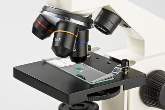
Is a microscope reflection or refraction? The underlying principal of a microscope is that lenses refract light which allows for magnification. … Taking advantage of the principle of refraction, devices can be built that can focus light. A device that produces converging or diverging light rays due to refraction is known as a lens.
Do microscopes use reflection or refraction? Microscopes use lenses that are responsible to attain the refraction of light of an object to visually magnify the image.
What are examples of reflection and refraction? Common objects include mirrors (reflect); glass of water with spoon in it (refract); foil (reflect); oil in a glass bottle (refract); prism (refract); glass (refract); lens (refract); or any shiny surface (reflect).
Is magnifying lens reflection or refraction? Magnifying glasses make objects appear larger because their convex lenses (convex means curved outward) refract or bend light rays, so that they converge or come together. … Despite the magnifying glass, your eyes trace the light rays back in parallel lines to the virtual image.
Is a microscope reflection or refraction? – Related Questions
What is the advantage of using a light microscope?
d. Advantage: Light microscopes have high magnification. Electron microscopes are helpful in viewing surface details of a specimen. Disadvantage: Light microscopes can be used only in the presence of light and have lower resolution.
How to focus a microscope correctly?
To focus a microscope, rotate to the lowest-power objective, and place your sample under the stage clips. Play with the magnification using the coarse adjustment knob and move your slide around until it is centered.
Why is depth of field important microscope?
Knowing the depth of field of the microscope at any given setting is important since it affects how much you have to move the specimen slide up, down, left, or right to image certain areas of the specimen, especially since it determines the required stability of the focusing axis.
What does simple squamous epithelium look like under a microscope?
A simple epithelium is only one layer of cells thick. … A squamous epithelial cell looks flat under a microscope. A cuboidal epithelial cell looks close to a square. A columnar epithelial cell looks like a column or a tall rectangle.
Are light microscopes?
The light microscope is an instrument for visualizing fine detail of an object. It does this by creating a magnified image through the use of a series of glass lenses, which first focus a beam of light onto or through an object, and convex objective lenses to enlarge the image formed.
How to use leeuwenhoek microscope?
Operation of the Leeuwenhoek microscope is simple. The specimen is placed on a pin that is manipulated by the means two of screws, one to adjust the distance between the specimen and lens and the other to adjust the height of the specimen.
What color is low power on a microscope?
Color the low-power objective purple. The high-power objective lens (H) has a magnification of 40X. Draw orange stripes on the high-power objective.
What kind of image does a scanning electron microscope make?
A scanning electron microscope (SEM) is a type of microscope which uses a focused beam of electrons to scan a surface of a sample to create a high resolution image. SEM produces images that can show information on a material’s surface composition and topography.
Are bacteria microscopic plants?
Bacteria are the smallest micro-organisms, ranging from between 0.0001 mm and 0.001 mm in size. Phytoplankton and protozoa range from about 0.001 mm to about 0.25 mm. The largest phytoplankton and protozoa can be seen with the naked eye, but most can only been seen under a microscope.
What is microscopic hair analysis?
Microscopic hair analysis is the science of comparing several strands of hair under a microscope and attempting to deduce if the strands ‘match’. It was accepted as a forensic science by the 1950s. Researchers often monitored more than a dozen attributes, including pigment distribution and scale patterns.
What is the highest power microscope lens?
The oil immersion objective lens provides the most powerful magnification, with a whopping magnification total of 1000x when combined with a 10x eyepiece.
How to prepare a specimen for a light microscope?
Preparation often involves nothing more than mounting a small piece of the specimen in a suitable liquid on a glass slide and covering it with a glass coverslip. ADVERTISEMENTS: The slide is then positioned on the specimen stage of the microscope and examined through the ocular lens, or with a camera.
Why must microscope be parcentric?
Overview. Parcentric and parfocal calibration compensate for the deviations from parfocality (focal plane) and parcentricity (collimation) that are normally encountered between different microscope objective lenses. They are both critical for maintaining proper position when changing magnification.
When to use a confocal microscope?
As a distinctive feature, confocal microscopy enables the creation of sharp images of the exact plane of focus, without any disturbing fluorescent light from the background or other regions of the specimen. Therefore, structures within thicker objects can be conveniently visualized using confocal microscopy.
Is microscopic colitis an inflammatory bowel disease?
Microscopic Colitis is an Inflammatory Bowel Disease (IBD) that affects the large bowel – the colon and rectum. It isn’t always as well-recognised as Crohn’s Disease or Ulcerative Colitis, other forms of IBD.
What microscope for micrometeorites?
A small and cheap microscope is enough to find the micrometeorites. It is important to use a wide-angle eyepiece and a low magnification. We use in this case an Omegon StereoView, 20x.
Who improved the first microscope?
It fell to a Dutch scientist, Anton van Leeuwenhoek, to make further improvements. Van Leeuwenhoek is sometimes popularly credited with the microscope’s invention.
What is low power objective in microscope?
Low power objectives cover a wide field of view and they are useful for examining large specimens or surveying many smaller specimens. This objective is useful for aligning the microscope. The power for the low objective is 10X.
How to adjust microscope focus?
Look at the objective lens (3) and the stage from the side and turn the focus knob (4) so the stage moves upward. Move it up as far as it will go without letting the objective touch the coverslip. Look through the eyepiece (1) and move the focus knob until the image comes into focus.
How to calculate field of view area microscope?
For instance, if your eyepiece reads 10X/22, and the magnification of your objective lens is 40. First, multiply 10 and 40 to get 400. Then divide 22 by 400 to get a FOV diameter of 0.055 millimeters.

