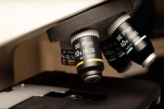
Is bright field microscope reflection or refraction? Brightfield microscopy uses transmitted light as a light source, compared to reflection contrast microscopy which uses incident light. The light passes through the specimen and is collected by the objective lens, which magnifies the light and further transmits it to an oracular lens through which the image is viewed.
Is a microscope reflection or refraction? The underlying principal of a microscope is that lenses refract light which allows for magnification. … Taking advantage of the principle of refraction, devices can be built that can focus light. A device that produces converging or diverging light rays due to refraction is known as a lens.
What is the principle of a bright field microscopy? For a specimen to be the focus and produce an image under the Brightfield Microscope, the specimen must pass through a uniform beam of the illuminating light. Through differential absorption and differential refraction, the microscope will produce a contrasting image.
How is reflection used in a microscope? In reflected light microscopy, illuminating light reaches the specimen, which may absorb some of the light and reflect some of the light, either in a specular or diffuse manner. Light that is returned upward can be captured by the objective in accordance with the objective’s numerical aperture.
Is bright field microscope reflection or refraction? – Related Questions
When did the light microscope invented?
In around 1590, Hans and Zacharias Janssen had created a microscope based on lenses in a tube [1]. No observations from these microscopes were published and it was not until Robert Hooke and Antonj van Leeuwenhoek that the microscope, as a scientific instrument, was born.
Can you see atoms in an electron microscope?
“So we can regularly see single atoms and atomic columns.” That’s because electron microscopes use a beam of electrons rather than photons, as you’d find in a regular light microscope. As electrons have a much shorter wavelength than photons, you can get much greater magnification and better resolution.
Who developed the microscope able to view cells?
1590: Two Dutch spectacle-makers and father-and-son team, Hans and Zacharias Janssen, create the first microscope. 1667: Robert Hooke’s famous “Micrographia” is published, which outlines Hooke’s various studies using the microscope.
When handling microscope slides?
Slides should be held by the edges, avoiding the cover glass area. Always begin viewing a slide using the microscope’s lowest magnification. This reduces the risk of contact by the microscope’s objective lens.
Can atoms be seen with a microscope?
Atoms are really small. So small, in fact, that it’s impossible to see one with the naked eye, even with the most powerful of microscopes. … Now, a photograph shows a single atom floating in an electric field, and it’s large enough to see without any kind of microscope.
Is ibs and microscopic colitis the same?
According to the review, the overlap between IBS and microscopic colitis differed based on study design. In case-control studies, microscopic colitis was seen more often in people who have IBS than those who had no symptoms.
What is compound microscope science definition?
A compound microscope is a microscope that uses multiple lenses to enlarge the image of a sample. … The total magnification is calculated by multiplying the magnification of the ocular lens by the magnification of the objective lens. Light is passed through the sample (called transmitted light illumination).
What type of microscope did leeuwenhoek use?
Antonie van Leeuwenhoek used single-lens microscopes, which he made, to make the first observations of bacteria and protozoa.
What does microscopic blood in your stool mean?
Blood may show up in your poop because of one or more of the following conditions: Growths or polyps of the colon. Hemorrhoids (swollen blood vessels near the anus and lower rectum that can rupture, causing bleeding) Anal fissures (splits or cracks in the lining of the anal opening)
How does microscopic agglutination test work?
The microscopic agglutination test (MAT) is the gold standard for sero-diagnosis of leptospirosis because of its unsurpassed diagnostic specificity. It uses panels of live leptospires, ideally recent isolates, representing the circulating serovars from the area where the patient became infected.
How do you focus the microscope?
To focus a microscope, rotate to the lowest-power objective, and place your sample under the stage clips. Play with the magnification using the coarse adjustment knob and move your slide around until it is centered.
Can you see nanoscale on a normal microscope?
In effect, many nanoscale objects are so small that light aimed at them misses, and so is not reflected back for us to see. This means that objects of less than 300 nm are distorted under a light microscope.
Can you see bacteria in water under a microscope?
In order to see bacteria, you will need to view them under the magnification of a microscopes as bacteria are too small to be observed by the naked eye. … At high magnification*, the bacterial cells will float in and out of focus, especially if the layer of water between the cover glass and the slide is too thick.
What is the purpose of diaphragm in microscope?
The field diaphragm controls how much light enters the substage condenser and, consequently, the rest of the microscope.
What does the light do on a microscope?
Light from a mirror is reflected up through the specimen, or object to be viewed, into the powerful objective lens, which produces the first magnification. The image produced by the objective lens is then magnified again by the eyepiece lens, which acts as a simple magnifying glass.
What kind of microscope to view rbc?
Description: This is a scanning electron microscope image from normal circulating human blood. One can see red blood cells, several white blood cells including lymphocytes, a monocyte, a neutrophil, and many small disc-shaped platelets.
When was the x ray microscope invented?
In 1948, Kirkpatrick and Baez (singer Joan Baez’s father) developed the first X-ray microscope. The early device had a resolution somewhere between the best optical and electron microscopes.
Why is a vacuum necessary in an electron microscope?
Most electron microscopes are high-vacuum instruments. Vacuums are needed to prevent electrical discharge in the gun assembly (arcing), and to allow the electrons to travel within the instrument unimpeded. … Also, any contaminants in the vacuum can be deposited upon the surface of the specimen as carbon.
Are omax microscopes good?
The OMAX has high quality objectives, excellent construction and design, lots of useful functions, accessories, customizable, and a lot of fun to use. If your looking to buy to buy a microscope to do college or professional work or use just looking to buy a hobby microscope look no further. The OMAX is for you.
Why are microscopes used?
A microscope is an instrument that is used to magnify small objects. Some microscopes can even be used to observe an object at the cellular level, allowing scientists to see the shape of a cell, its nucleus, mitochondria, and other organelles.
How to hold microscope properly?
Always carry the microscope with 2 hands—place one hand on the microscope arm and the other hand under the microscope base. Do not touch the objective lenses (i.e. the tips of the objectives). Keep the objectives in the scan position and keep the stage low when adding or removing slides.

