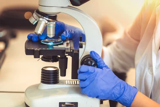
Is higher power lens longer microscope? Going to high power on a microscope decreases the area of the field of view. The field of view is inversely proportional to the magnification of the objective lens. … The specimen appears larger with a higher magnification because a smaller area of the object is spread out to cover the field of view of your eye.
Is the high power objective lens the longest? Objective Lenses: Usually you will find 3 or 4 objective lenses on a microscope. They almost always consist of 4X, 10X, 40X and 100X powers. … The shortest lens is the lowest power, the longest one is the lens with the greatest power. The high power objective lenses are retractable (i.e. 40XR).
What is the difference between 4X 10x and 40x on a microscope? For example, optical (light) microscopes are usually equipped with four objectives: 4x and 10x are low power objectives; 40x and 100õ are powerful ones. The total magnification (received with 10x eyepiece) of less than 400x characterizes the microscope as a low-powered model; more than 400x as a powerful one.
What is higher power on a microscope? When you switch to a higher power, the field of view is closes in. You will see more of an object on low power. The depth of focus is greatest on the lowest power objective. Each time you switch to a higher power, the depth of focus is reduced. Therefore a smaller part of the specimen is in focus at higher power.
Is higher power lens longer microscope? – Related Questions
What does microscopic haematuria mean?
Microscopic hematuria means that the blood can only be seen with a microscope. Gross hematuria means the urine appears red or the color of tea or cola to the naked eye.
What the first microscope look like?
The early simple “microscopes” which were really only magnifying glasses had one power, usually about 6X – 10X . One thing that was very common and interesting to look at was fleas and other tiny insects. These early magnifiers were hence called “flea glasses”.
Can a light microscope see living cell?
There are two main types of microscope: light microscopes are used to study living cells and for regular use when relatively low magnification and resolution is enough. electron microscopes provide higher magnifications and higher resolution images but cannot be used to view living cells.
What is the eyepiece on a microscope called?
The eyepiece, or ocular lens, is the part of the microscope that magnifies the image produced by the microscope’s objective so that it can be seen by the human eye.
Are quantum principles microscopic?
Quantum microscopy allows microscopic properties of matter and quantum particles to be measured and imaged. Various types of microscopy use quantum principles.
What does microscopic mean in greek?
The word is a scientific term if you literally mean “can be seen with a microscope,” although people use it sometimes to mean “really small,” as in the phrase “Wow, your feet are microscopic.” Mikros means “small” in Greek, and the scope part of the word comes from the Greek word skopein, “to examine.”
Can uterine fibroids cause microscopic blood in urine?
If any blood appears in your bladder, it can be the sign of large fibroids. These cases are not common, but there have been cases where the bladder can become damaged from uterine fibroids. If one of these growths causes a rip in the bladder, it can cause leakage of urine throughout the body, which has a fatal outcome.
How to adjust brightness on a microscope?
Brightness is related to the illumination system and can be changed by changing the voltage to the lamp (rheostat) and adjusting the condenser and diaphragm/pinhole apertures. Brightness is also related to the numerical aperture of the objective lens (the larger the numerical aperture, the brighter the image).
What is opmi microscope?
ZEISS OPMI Sensera is an ideal surgical microscope for laser microsurgery, especially when equipped with the optionally available external fine focusing module, which keeps the laser and microscope focus synchronized during surgery.
What is numerical aperture of compound microscope?
The numerical aperture of a microscope objective is the measure of its ability to gather light and to resolve fine specimen detail while working at a fixed object (or specimen) distance.
What is the magnification range of a tem microscope?
Transmission electron microscopes (TEM) are microscopes that use a particle beam of electrons to visualize specimens and generate a highly-magnified image. TEMs can magnify objects up to 2 million times.
How microscopes help us today?
A microscope lets the user see the tiniest parts of our world: microbes, small structures within larger objects and even the molecules that are the building blocks of all matter. The ability to see otherwise invisible things enriches our lives on many levels.
Is a virus microscopic or macroscopic?
Viruses are microscopic parasites, generally much smaller than bacteria. They lack the capacity to thrive and reproduce outside of a host body.
What level of magnification is microscopic?
Calculate total magnification by multiplying the eyepiece magnification by the objective lens magnification. Typical laboratory microscopes magnify objects 40x, 100x and 400x.
Why are light microscopes so useful in biology?
light microscopes are used to study living cells and for regular use when relatively low magnification and resolution is enough. electron microscopes provide higher magnifications and higher resolution images but cannot be used to view living cells.
How would a letter e look like under a microscope?
The letter “e” appears upside down and backwards under a microscope. Either, diatoms are single celled, or they do not have a cell wall.
How often should you clean the lenses of your microscope?
The microscope stage is cleaned in a similar manner to the body tube, first with a moistened cloth, then with a dry one. Because of the variety of contaminants that may be deposited on the stage from specimens and from constant handling and manipulation, it should be cleaned after every use of the microscope.
How is the image produced in a light microscope?
The light microscope is an instrument for visualizing fine detail of an object. It does this by creating a magnified image through the use of a series of glass lenses, which first focus a beam of light onto or through an object, and convex objective lenses to enlarge the image formed.
What is the smallest thing an electron microscope can see?
Light microscopes let us look at objects as long as a millimetre (10-3 m) and as small as 0.2 micrometres (0.2 thousands of a millimetre or 2 x 10⁻⁷ m), whereas the most powerful electron microscopes allow us to see objects as small as an atom (about one ten-millionth of a millimetre or 1 angstrom or 10-10 m).
Is microscopic blood in urine a sign of bladder cancer?
In most cases, blood in the urine (called hematuria) is the first sign of bladder cancer. There may be enough blood to change the color of the urine to orange, pink, or, less often, dark red.
What is viewed on a two photon microscope?
Two photon microscopes can look deep into fluorescent samples, typically 5-20 times deeper than other types of fluorescent microscopes. Our 2-P microscope can image four simultaneous color channels at 30 frames/second. It is well automated with computer control of the focus, stage motion and timing.

