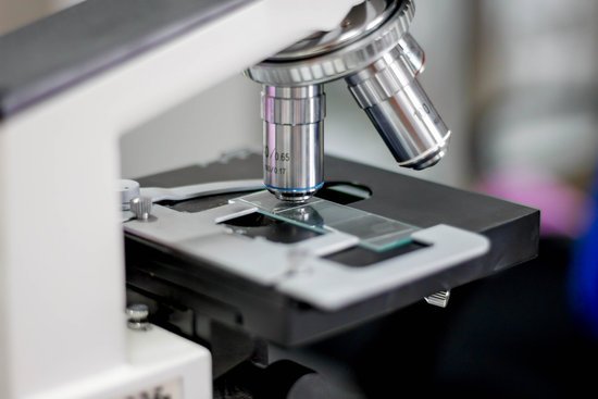
Is microscope parfocal? A. Parfocal means that the microscope is binocular. … Parfocal means that when one objective lens is in focus, then the other objectives will also be in focus.
How can you tell if a microscope is parfocal? To determine if a microscope has parfocal objectives, a slide should be brought into focus using the highest magnification settings. The operator should then switch to an objective with a lower magnification level to check for sharpness of focus on the slide.
Are all microscopes parfocal? Microscopy. Parfocal microscope objectives stay in focus when magnification is changed; i.e., if the microscope is switched from a lower power objective (e.g., 10×) to a higher power objective (e.g., 40×), the object stays in focus. Most modern bright-field microscopes are parfocal.
What is conventional light microscopy? The conventional microscope uses visible light (400-700 nanometers) to illuminate and produce a magnified image of a sample. … This fluorescent species in turn emits a lower energy light of a longer wavelength that produces the magnified image instead of the original light source.
Is microscope parfocal? – Related Questions
Can you view living things with an electron microscope?
Electron microscopes are the most powerful type of microscope, capable of distinguishing even individual atoms. However, these microscopes cannot be used to image living cells because the electrons destroy the samples.
How is a parfocal microscope helpful?
It is helpful for a microscope to be parfocal because the user does not have to adjust the focus when changing the power of magnification.
How do you calculate magnification on a compound microscope?
To calculate the total magnification of the compound light microscope multiply the magnification power of the ocular lens by the power of the objective lens. For instance, a 10x ocular and a 40x objective would have a 400x total magnification. The highest total magnification for a compound light microscope is 1000x.
What is the eyepiece lens of a microscope called?
An eyepiece, or ocular lens, is a type of lens that is attached to a variety of optical devices such as telescopes and microscopes. It is so named because it is usually the lens that is closest to the eye when someone looks through the device.
Which type of microscope has the highest magnification?
Out of all types of microscopes, the electron microscope has the greatest capability in achieving high magnification and resolution levels, enabling us to look at things right down to each individual atom.
What does the inclination joint do on a microscope?
Inclination Joint: Where the microscope arm connects to the microscope base, there may be a pin. If so, you can place one hand on the base and with the other hand grab the arm and rotate it back. It will tilt your microscope back for more comfortable viewing.
How image is formed in microscope?
Image formation in a microscope, according to the Abbe theory. Specimens are illuminated by light from a condenser. … The microscope objective collects these diffracted waves and directs them to the focal plane, where interference between the diffracted waves produces an image of the object.
How image formed in microscope?
Section Overview: In the optical microscope, image formation occurs at the intermediate image plane through interference between direct light that has passed through the specimen unaltered and light diffracted by minute features present in the specimen.
Can nsaids cause microscopic hematuria?
Yes, ibuprofen can cause hematuria (blood in the urine). Due to you having blood in your urine it would most likely be recommended that you do not take ibuprofen or other NSAID in the future, unless you have been prescribed them. Other non-steroidal anti-inflammatory (NSAID) drugs may cause the same side effect.
How to clean a microscope inside eyepiece?
To clean the eyepiece lens of your microscope, breathe onto the eyepiece lens and then wipe with lens tissue. For dirt that is difficult to remove, add ethanol (methanol in extreme cases) to a cotton swab, wipe the surface and then dry with a dry swab.
What is the use of condenser in microscope?
On upright microscopes, the condenser is located beneath the stage and serves to gather wavefronts from the microscope light source and concentrate them into a cone of light that illuminates the specimen with uniform intensity over the entire viewfield.
What regulates the amount of light entering the microscope?
The condenser is equipped with an iris diaphragm, a shutter controlled by a lever that is used to regulate the amount of light entering the lens system. Above the stage and attached to the arm of the microscope is the body tube. This structure houses the lens system that magnifies the specimen.
What is the maximum total magnification for a light microscope?
Using the mathematical equations given above and the values for maximum numerical aperture attainable with the lenses of a light microscope it can be shown that the maximum useful magnification on a light microscope is between 1000X and 1500X. Higher magnification is possible, but resolution will not improve.
Do light microscopes use lenses?
The light microscope is an instrument for visualizing fine detail of an object. It does this by creating a magnified image through the use of a series of glass lenses, which first focus a beam of light onto or through an object, and convex objective lenses to enlarge the image formed.
How can microscopes be used in the field of science?
A microscope is an instrument that is used to magnify small objects. Some microscopes can even be used to observe an object at the cellular level, allowing scientists to see the shape of a cell, its nucleus, mitochondria, and other organelles.
How did zacharias janssen invent the microscope?
A Dutch father-son team named Hans and Zacharias Janssen invented the first so-called compound microscope in the late 16th century when they discovered that, if they put a lens at the top and bottom of a tube and looked through it, objects on the other end became magnified.
Can you see atoms under a microscope?
Atoms are really small. So small, in fact, that it’s impossible to see one with the naked eye, even with the most powerful of microscopes. … Now, a photograph shows a single atom floating in an electric field, and it’s large enough to see without any kind of microscope.
How to view mold under microscope?
Place a drop of water in the center of the slide, using an eyedropper if you have one, or the tip of a clean finger. You can use solution of methylene blue instead, which is a microscope stain, and makes the sample easier to see by coloring certain parts of the mold cells.
What is the basic microscopic anatomy of cardiac muscle?
Cardiac muscles are composed of tubular cardiomyocytes, or cardiac muscle cells. The cardiomyocytes are composed of tubular myofibrils, which are repeating sections of sarcomeres. Intercalated disks transmit electrical action potentials between sarcomeres.
How to account for magnification on a microscope?
To figure the total magnification of an image that you are viewing through the microscope is really quite simple. To get the total magnification take the power of the objective (4X, 10X, 40x) and multiply by the power of the eyepiece, usually 10X.

