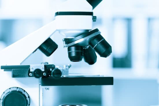
Is there a specific type of oil for a microscope? Immersion oils are transparent oils that have specific optical and viscosity characteristics necessary for use in microscopy. Typical oils used have an index of refraction of around 1.515.
What type of oil is used under the 100x objective? Why do you use immersion oil with 100x objective lens quizlet? Immersion oil has the same refractive index compared with that of glass. This prevents light loss due to diffraction. Oil immersion should be used between the slide and 100x objective lens, this is a special oil that has the same refractive index as glass.
Why cedar oil is used in microscope? By placing a substance such as immersion oil with a refractive index equal to that of the glass slide in the space filled with air, more light is directed through the objective and a clearer image is observed.
What is microscope immersion oil? Immersion oil increases the resolving power of the microscope by replacing the air gap between the immersion objective lens and cover glass with a high refractive index medium and reducing light refraction. Nikon manufactures four types of Immersion Oil for microscopy.
Is there a specific type of oil for a microscope? – Related Questions
What does the base on a microscope?
Base: The bottom of the microscope, used for support. … If your microscope has a mirror, it is used to reflect light from an external light source up through the bottom of the stage.
Do probiotics help microscopic colitis?
Some researchers have suggested that probiotics may benefit people with MC because these bacteria and yeasts can help relieve symptoms of other gut conditions, such as irritable bowel syndrome (IBS) and ulcerative colitis.
What is the difference between macroscopic and microscopic kinetic energy?
Kinetic energy is the energy of motion. This can be the motion of large objects (macroscopic kinetic energy), or the movement of small atoms and molecules (microscopic kinetic energy). Macroscopic kinetic energy is “high quality” energy, while microscopic kinetic energy is more disordered and “low-quality.”
How to calculate the ocular power of a microscope?
To calculate the total magnification of the compound light microscope multiply the magnification power of the ocular lens by the power of the objective lens. For instance, a 10x ocular and a 40x objective would have a 400x total magnification. The highest total magnification for a compound light microscope is 1000x.
What image does a light microscope produce?
Principles. The light microscope is an instrument for visualizing fine detail of an object. It does this by creating a magnified image through the use of a series of glass lenses, which first focus a beam of light onto or through an object, and convex objective lenses to enlarge the image formed.
What microscope would you use to view bordetella pertussis?
A frequently utilized tool is Confocal Laser Scanning Microscopy. We present a detailed protocol to grow, observe and analyze biofilms of the respiratory human pathogen, Bordetella pertussis in space and time.
Why do scientists use electron microscopes?
The electron microscope is an integral part of many laboratories. Researchers use it to examine biological materials (such as microorganisms and cells), a variety of large molecules, medical biopsy samples, metals and crystalline structures, and the characteristics of various surfaces.
How do i calculate magnification on a microscope?
It’s very easy to figure out the magnification of your microscope. Simply multiply the magnification of the eyepiece by the magnification of the objective lens. The magnification of both microscope eyepieces and objectives is almost always engraved on the barrel (objective) or top (eyepiece).
What regulates the amount of light in the microscope?
The condenser is equipped with an iris diaphragm, a shutter controlled by a lever that is used to regulate the amount of light entering the lens system. Above the stage and attached to the arm of the microscope is the body tube. This structure houses the lens system that magnifies the specimen.
What are the microscopic organisms in the ocean?
The four main types of micro-organisms in the ocean are: Algae — these are single celled plants also known as phytoplankton (from the Greek, meaning drifting plants).
When viewed under a microscope gram positive bacteria?
Gram positive bacteria have a distinctive purple appearance when observed under a light microscope following Gram staining. This is due to retention of the purple crystal violet stain in the thick peptidoglycan layer of the cell wall.
What is the advantage of scanning electron microscope?
Advantages of a Scanning Electron Microscope include its wide-array of applications, the detailed three-dimensional and topographical imaging and the versatile information garnered from different detectors.
What is fluorescence and its advantage in microscope imaging?
Fluorescence optical microscopy is a powerful imaging tool in biology used to collect spatial and functional in- formation about both endogenous autofluorescent and exogenously labeled molecules and structures. Fluores- cent molecules enable researchers to obtain spatial and functional information.
How did the microscope impact the renaissance?
The development of the microscope during the European Renaissance impacted both the ancient world and modern world by giving students something to learn in the classrooms and other scientists things to discover. Around year 1595 the microscope, one of the greatest inventions, was made.
What is the purpose of a compound microscope?
Typically, a compound microscope is used for viewing samples at high magnification (40 – 1000x), which is achieved by the combined effect of two sets of lenses: the ocular lens (in the eyepiece) and the objective lenses (close to the sample).
What was the microscope used for?
A microscope is an instrument that can be used to observe small objects, even cells. The image of an object is magnified through at least one lens in the microscope. This lens bends light toward the eye and makes an object appear larger than it actually is.
What is hemocytometer microscope?
The hemocytometer (or haemocytometer) is a counting-chamber device originally designed and usually used for counting blood cells. The hemocytometer was invented by Louis-Charles Malassez and consists of a thick glass microscope slide with a rectangular indentation that creates a precision volume chamber.
Why do we use microscopes to study cells?
A cell is the smallest unit of life. Most cells are so small that they cannot be viewed with the naked eye. Therefore, scientists must use microscopes to study cells. Electron microscopes provide higher magnification, higher resolution, and more detail than light microscopes.
Can advil cause microscopic hematuria?
Yes, ibuprofen can cause hematuria (blood in the urine). Due to you having blood in your urine it would most likely be recommended that you do not take ibuprofen or other NSAID in the future, unless you have been prescribed them.
Can i see plant cells in a microscope?
Thus, most cells in their natural state, even if fixed and sectioned, are almost invisible in an ordinary light microscope. One way to make them visible is to stain them with dyes.
How were microscopes made?
A Dutch father-son team named Hans and Zacharias Janssen invented the first so-called compound microscope in the late 16th century when they discovered that, if they put a lens at the top and bottom of a tube and looked through it, objects on the other end became magnified.

