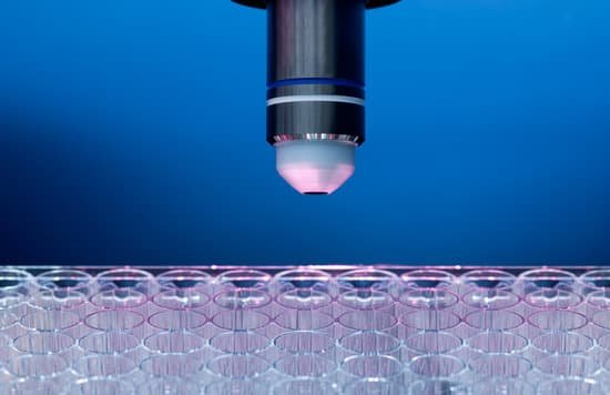
What are microscopes and their uses? A microscope, from the Ancient Greek words mikrós or “small” and skopeîn or “to look or see,” is a tool that is used to view smaller objects that the human eye can see. Microscopy is the scientific field of study which is used to study minute structures and objects by a microscope.
What is microscope and its uses? A microscope is an instrument that can be used to observe small objects, even cells. The image of an object is magnified through at least one lens in the microscope. This lens bends light toward the eye and makes an object appear larger than it actually is.
What are the 5 uses of microscope? There are three basic types of microscopes: optical, charged particle (electron and ion), and scanning probe. Optical microscopes are the ones most familiar to everyone from the high school science lab or the doctor’s office.
What are the 3 types of microscope? Microscopes are a mainstay in life science research but advances in imaging have allowed their use to expand into most areas of science and technology. They are commonly used to view different types of cells, analyze clinical specimens and to scan nanomaterials.
What are microscopes and their uses? – Related Questions
How to use usb microscope camera?
Plug the device into any open USB port on the computer or the television. Hold the microscope and lightly touch the lens to the specimen. The image should now be visible on the monitor or television screen. These microscopes should only be used to examine dry specimens.
How to use a microscope micrometer?
Procedure. Place a stage micrometer on the microscope stage, and using the lowest magnification (4X), focus on the grid of the stage micrometer. Rotate the ocular micrometer by turning the appropriate eyepiece. Move the stage until you superimpose the lines of the ocular micrometer upon those of the stage micrometer.
What is the highest objective on a microscope?
The high-powered objective lens (also called “high dry” lens) is ideal for observing fine details within a specimen sample.
How did the electron microscope work?
The electron microscope uses a beam of electrons and their wave-like characteristics to magnify an object’s image, unlike the optical microscope that uses visible light to magnify images. … These interactions and effects are detected and transformed into an image.
How did the development of microscopes?
In the late 16th century several Dutch lens makers designed devices that magnified objects, but in 1609 Galileo Galilei perfected the first device known as a microscope. Dutch spectacle makers Zaccharias Janssen and Hans Lipperhey are noted as the first men to develop the concept of the compound microscope.
How to sit at a microscope?
Adopt a correct sitting posture. For forward activities such as using a microscope, it is best to sit with an open hip angle, i.e. with your upper legs at an angle of at least 135° to your torso. Make sure that your work surface is at elbow height. A mechanically or electrically adjustable lab table is ideal.
What is an slides on a microscope?
A microscope slide is a thin flat piece of glass, typically 75 by 26 mm (3 by 1 inches) and about 1 mm thick, used to hold objects for examination under a microscope. Typically the object is mounted (secured) on the slide, and then both are inserted together in the microscope for viewing.
When was the microscope discovered?
In around 1590, Hans and Zacharias Janssen had created a microscope based on lenses in a tube [1]. No observations from these microscopes were published and it was not until Robert Hooke and Antonj van Leeuwenhoek that the microscope, as a scientific instrument, was born.
What is the highest magnification microscope?
The microscope that can achieve the highest magnification and greatest resolution is the electron microscope, which is an optical instrument that is designed to enable us to see microscopic details down to the atomic scale (check also atom microscopy).
What is the most powerful light microscope?
Lawrence Berkeley National Labs just turned on a $27 million electron microscope. Its ability to make images to a resolution of half the width of a hydrogen atom makes it the most powerful microscope in the world.
Why is the metallurgy microscope different from biological?
A simple answer is that a metallurgical microscope is another type of light microscopes and unlike a biological microscope it uses a reflected white light. Obviously the nature of samples is different for such a use, i.e. a metal, semiconductor or plastic rather than a biology slide, cell, living microorganism etc.
When using a compound microscope the areas above and below?
When using a compound microscope, the areas above and below the depth of focus are best viewed by adjusting the: fine or coarse adjustment. Which microscope has the largest potential working distance?
What does eosinophil look like under a microscope?
Eosinophils contain large granules, and the nucleus exists as two nonsegmented lobes. In addition, the granules of eosinophils typically stain red, which makes them easily distinguished from other granulocytes when viewed on prepared slides under a microscope.
What can be seen with a transmission electron microscope?
The transmission electron microscope is used to view thin specimens (tissue sections, molecules, etc) through which electrons can pass generating a projection image. The TEM is analogous in many ways to the conventional (compound) light microscope.
How have electron microscopes helped scientists understand cells?
The development of the electron microscopes therefore helped scientists to learn about the sub-cellular structures involved in aerobic respiration called mitochondria . The scientists developed their explanations about how the structure of the mitochondria allowed it to efficiently carry out aerobic respiration.
How to calculate smallest thing under a microscope?
λ/2 where d is limit of resolution and λ is the wavelength of the type of wave/particle you are using in your microscope.
What makes something microscopic?
When people say “microscopic” they mean that the thing or the event is so small you cannot observe it properly without some sort of aid, such as a microscope. … Essentially, it described objects and events that are so small we do not measure or directly observe the precise state of a thermodynamic system.
How microscope magnify an object?
A microscope is an instrument that can be used to observe small objects, even cells. The image of an object is magnified through at least one lens in the microscope. This lens bends light toward the eye and makes an object appear larger than it actually is.
What do compound microscopes do?
Typically, a compound microscope is used for viewing samples at high magnification (40 – 1000x), which is achieved by the combined effect of two sets of lenses: the ocular lens (in the eyepiece) and the objective lenses (close to the sample).
Can microscopic pieces of glass hurt you if consumed?
Sharp objects can become stuck and lead to a puncture in the digestive tract. Small pieces of glass generally pass without any symptoms.
When did janssen invent the microscope?
Lens Crafters Circa 1590: Invention of the Microscope. Every major field of science has benefited from the use of some form of microscope, an invention that dates back to the late 16th century and a modest Dutch eyeglass maker named Zacharias Janssen.

