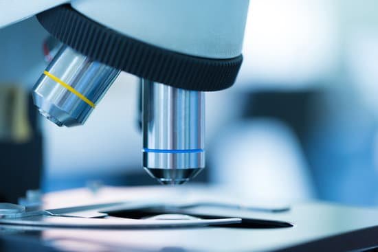
What are transmission electron microscopes used to look at? The transmission electron microscope is used to view thin specimens (tissue sections, molecules, etc) through which electrons can pass generating a projection image. The TEM is analogous in many ways to the conventional (compound) light microscope.
What can TEM measure? TEM is a well-known imaging technique that uses beam of electrons as seen in Fig. 5.7. … While TEM is used to measure the particle size of QDs, it can also provide information about the size distribution of the corresponding QD materials.
What type of information can be retrieved from transmission electron microscope? TEM can be used to study the growth of layers, their composition and defects in semiconductors. High resolution can be used to analyze the quality, shape, size and density of quantum wells, wires and dots. The TEM operates on the same basic principles as the light microscope but uses electrons instead of light.
Who invented microscope today? Zacharias Janssen, credited with inventing the microscope. (Image credit: Public domain.) For millennia, the smallest thing humans could see was about as wide as a human hair. When the microscope was invented around 1590, suddenly we saw a new world of living things in our water, in our food and under our nose.
What are transmission electron microscopes used to look at? – Related Questions
What is microscopic anatomy called?
Microscopic anatomy: The study of normal structure of an organism under the microscope. Known among medical students simply as ‘micro. ‘ Also known as histology.
Can you see lyme with a microscope?
It can be detected by light microscopy in tissue sections or, rarely, in blood smears using various staining methods.
What type of microscope is needed to see small molecules?
Some electron microscopes can detect objects that are approximately one-twentieth of a nanometre (10-9 m) in size – they can be used to visualise objects as small as viruses, molecules or even individual atoms.
Who pioneered the use of microscope?
Grinding glass to use for spectacles and magnifying glasses was commonplace during the 13th century. In the late 16th century several Dutch lens makers designed devices that magnified objects, but in 1609 Galileo Galilei perfected the first device known as a microscope.
How does a microscope magnify quizlet?
The number of times larger an image appears compared with the actual size of the object. Microscopes produce linear magnification, meaning that if an object is magnified by x100, it appears 100 times larger. How do you calculate total magnification in optical microscopes? You just studied 22 terms!
How do you focus an electron microscope?
Electron microscopes use a beam of electrons rather than visible light to illuminate the sample. They focus the electron beam using electromagnetic coils instead of glass lenses (as a light microscope does) because electrons can’t pass through glass.
Why can you see dna without a microscope?
When molecules are insoluble (unable to be dissolved), they clump together and become visible. DNA is not soluble in alcohol; therefore, it makes the DNA strands clump together and become visible to the naked eye.
What is liquid lens microscope?
Focus stacking, or z-stacking, is the combining of images taken at different focus distances. Liquid lenses can be integrated into tube lenses or in infinity space within a microscope to quickly and precisely focus to various object planes.
How small can electron microscope see?
Light microscopes let us look at objects as long as a millimetre (10-3 m) and as small as 0.2 micrometres (0.2 thousands of a millimetre or 2 x 10⁻⁷ m), whereas the most powerful electron microscopes allow us to see objects as small as an atom (about one ten-millionth of a millimetre or 1 angstrom or 10-10 m).
What field of vision refers to microscope?
Introduction. Microscope field of view (FOV) is the maximum area visible when looking through the microscope eyepiece (eyepiece FOV) or scientific camera (camera FOV), usually quoted as a diameter measurement (Figure 1).
What is the objective magnification of a microscope?
Objectives typically have magnifying powers that range from 1:1 (1X) to 100:1 (100X), with the most common powers being 4X (or 5X), 10X, 20X, 40X (or 50X), and 100X.
What is the glass that is used with microscopes?
A microscope slide is a thin flat piece of glass, typically 75 by 26 mm (3 by 1 inches) and about 1 mm thick, used to hold objects for examination under a microscope.
Why are viruses not resolved by a light compound microscope?
Standard light microscopes allow us to see our cells clearly. However, these microscopes are limited by light itself as they cannot show anything smaller than half the wavelength of visible light – and viruses are much smaller than this. But we can use microscopes to see the damage viruses do to our cells.
What property of a microscope image is described by magnification?
In addition, objective magnification also plays a role in determining image brightness, which is inversely proportional to the square of the lateral magnification. The square of the numerical aperture/magnification ratio expresses the light-gathering power of the objective when utilized with transmitted illumination.
How are compound microscopes used?
Typically, a compound microscope is used for viewing samples at high magnification (40 – 1000x), which is achieved by the combined effect of two sets of lenses: the ocular lens (in the eyepiece) and the objective lenses (close to the sample).
How to use microscope on computer?
Plug the device into any open USB port on the computer or the television. Hold the microscope and lightly touch the lens to the specimen. The image should now be visible on the monitor or television screen. These microscopes should only be used to examine dry specimens.
How to work out microscope magnification?
To calculate the total magnification of the compound light microscope multiply the magnification power of the ocular lens by the power of the objective lens. For instance, a 10x ocular and a 40x objective would have a 400x total magnification. The highest total magnification for a compound light microscope is 1000x.
Do nurses use microscopes?
The operating room (OR) nurse plays a crucial role, contributing to a smooth surgical procedure. … 20 of these operating nurses are specialized in ENT and work with surgical microscopes on a daily basis.
Which part of the microscope do you look through?
Typically, a compound microscope has one lens in the eyepiece, the part you look through. The eyepiece lens usually magnifies 10 .
What are the types of optical microscope?
There are two basic types of optical microscopes: simple microscopes and compound microscopes. A simple microscope uses the optical power of single lens or group of lenses for magnification.

