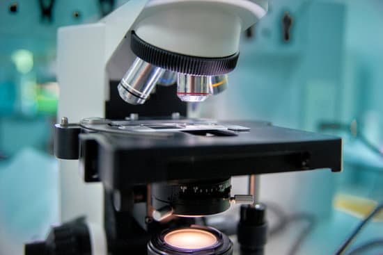
What can you see in a light microscope? Explanation: You can see most bacteria and some organelles like mitochondria plus the human egg. You can not see the very smallest bacteria, viruses, macromolecules, ribosomes, proteins, and of course atoms.
What does a light microscope allow you to see? Principles. The light microscope is an instrument for visualizing fine detail of an object. It does this by creating a magnified image through the use of a series of glass lenses, which first focus a beam of light onto or through an object, and convex objective lenses to enlarge the image formed.
What does a light microscope see an image? It is through the microscope’s lenses that the image of an object can be magnified and observed in detail. A simple light microscope manipulates how light enters the eye using a convex lens, where both sides of the lens are curved outwards.
What cell structure can be seen with a light microscope? Note: The nucleus, cytoplasm, cell membrane, chloroplasts and cell wall are organelles which can be seen under a light microscope.
What can you see in a light microscope? – Related Questions
Why do we use microscopes?
A microscope is an instrument that is used to magnify small objects. Some microscopes can even be used to observe an object at the cellular level, allowing scientists to see the shape of a cell, its nucleus, mitochondria, and other organelles.
Where is the phase plate located in the microscope?
A plate that causes a change in the phase of an electron wave. The phase plate placed at the back focal plane of an electron microscope creates a relative phase change between the transmitted wave and scattered waves from a specimen.
What is the maximum magnification of a dissecting microscope?
A dissecting microscope is used to view three-dimensional objects and larger specimens, with a maximum magnification of 100x. This type of microscope might be used to study external features on an object or to examine structures not easily mounted onto flat slides. Both microscopes have similar features.
What are the two types of microscopic colitis?
Two types of microscopic colitis are lymphocytic colitis and collagenous colitis. The two types cause different changes in colon tissue. In lymphocytic colitis, the colon lining contains more white blood cells than normal.
What is a low mag microscope called?
The stereo- or dissecting microscope is an optical microscope variant designed for observation with low magnification (2 – 100x) using incident light illumination (light reflected off the surface of the sample is observed by the user), although it can also be combined with transmitted light in some instruments.
What are the magnifications of the three microscope objectives?
Objective lenses come in various magnification powers, with the most common being 4x, 10x, 40x, and 100x, also known as scanning, low power, high power, and (typically) oil immersion objectives, respectively.
What is the diaphragm used for in a microscope?
Opening and closing of the condenser aperture diaphragm controls the angle of the light cone reaching the specimen. The setting of the condenser’s aperture diaphragm, along with the aperture of the objective, determines the realized numerical aperture of the microscope system.
How have microscopes impacted on human health?
The microscope has had a major impact in the medical field. Doctors use microscopes to spot abnormal cells and to identify the different types of cells. This helps in identifying and treating diseases such as sickle cell caused by abnormal cells that have a sickle like shape.
Who discovered electron microscope in 1940?
1940: Vladimir Zworykin, better known as a co-inventor of television, demonstrates the first electron microscope in the United States.
How to clean the microscope lens?
Dip a lens wipe or cotton swab into distilled water and shake off any excess liquid. Then, wipe the lens using the spiral motion. This should remove all water-soluble dirt.
Which microscope is used to see cells?
Two types of electron microscopy—transmission and scanning—are widely used to study cells. In principle, transmission electron microscopy is similar to the observation of stained cells with the bright-field light microscope.
How can acquiring charge be described at a microscopic level?
How can acquiring charge be described at a microscopic level? It is a process of balancing the charge on an atom. It is the process of creating charge.
What is resolving power of compound microscope?
The resolving power is the capacity of an instrument to resolve two points that are close together. The resolving power of a compound microscope is 0.25μm.
Can i look at bacteria via microscope petri dish?
Viewing bacteria under a microscope is much the same as looking at anything under a microscope. Prepare the sample of bacteria on a slide and place under the microscope on the stage. Adjust the focus then change the objective lens until the bacteria come into the field of view.
How to see sperm with a microscope?
You can view sperm at 400x magnification. You do NOT want a microscope that advertises anything above 1000x, it is just empty magnification and is unnecessary. In order to examine semen with the microscope you will need depression slides, cover slips, and a biological microscope.
What is meant by a compound light microscope?
A compound light microscope is a microscope with more than one lens and its own light source. In this type of microscope, there are ocular lenses in the binocular eyepieces and objective lenses in a rotating nosepiece closer to the specimen.
Which of the following microscopes uses visible light?
The optical microscope, often referred to as the “light optical microscope,” is a type of microscope that uses visible light and a system of lenses to magnify images of small samples.
Can you see proton with electron microscope?
We can never see the subatomic particles directly, but can only infer from observation of such indirect effects like tracks. If there are many of them and they are emitting some radiation, and also if we shine some radiation on then and receive back the response this will also constitute a kind of seeing.
How does a compound microscope work in general?
A compound microscope uses two or more lenses to produce a magnified image of an object, known as a specimen, placed on a slide (a piece of glass) at the base. … By raising and lowering the stage, you move the lenses closer to or further away from the object you’re examining, adjusting the focus of the image you see.
Are microscopes bad for your eyes?
The narrow field of view from most microscope eyepieces is a major cause of eye strain and bad posture. Users who wear spectacles often have to remove them, increasing the risk of eye strain; and many users also suffer the distraction of floating fragments of tissue debris in the eye.
Why would there be microscopic blood in urine?
Microscopic urinary bleeding is a common symptom of glomerulonephritis, an inflammation of the kidneys’ filtering system. Glomerulonephritis may be part of a systemic disease, such as diabetes, or it can occur on its own.

